EFA Medical Conference 2017
"Breast, Ovary and Endometriosis"
October 28, 2017 - Lotte New York Palace Hotel
Breast Cancer in Young Women; Early Diagnosis Timely Intervention
Stephanie Bernik, MD, FACS
Thank you for having me. My talk is going to be focused on breast cancer in young women. Some of this has been covered. Some of it will also be touched on later in the talk, but I will try and go through this in an educational way, and I'll try to speed it up a little bit.
In general, I'm going to talk about some advances in radiation. We'll talk about some reconstructive advances. There's a talk later on in the day with Dr. [Orin Lehrman 00:00:41]. He is my partner in crime at Lenox Hill. We have improved screening techniques, which I'll discuss. We'll talk about breast conservation and some of the advances that we have in the present day. Chemoprevention will be discussed, and, obviously, that touches on estrogen in breast cancer. And we'll talk about some of the changes that have occurred with the use of the sentinel node biopsy.
In general, we already heard some of this. There are about 240,000 cases of breast cancer a year. 12,000 of these are in women under the age of 40, and 26,000 are under the age of 45, so this does affect a good proportion of young women, although it's a minority of the cases of breast cancer. 1,000 women under the age of 40 die of breast cancer every year, and these are women that have been young children, are in their most productive years of their life, so it really is a tragedy. Because we don't have routine screening for women under the age of 40, 80 percent of these women coming in have felt a mass. We also do see breast cancer in pregnancy. It occurs in about 1 in 3,000 pregnancies.
Although most tumors in young women are estrogen receptor positive, there's less of a proportion of estrogen receptor positive cancers in young women. We're seeing more estrogen receptor negative tumors in younger women. Tumors are usually of a higher grade and a larger size. Some of that goes back to the fact that we're not routinely screening these women, so they're coming in when you can feel a mass. Certainly, when you feel a mass, it's bigger than a mass you would see on one of the screening methods we standardly use in women that are older. Some studies do suggest, even if we catch the tumor at the same stage as any younger woman compared to that of an older woman, that the outcome will be worse. The reasons for this are unclear, but it may be that there is increased circulating estrogen in a young woman's body.
Here, you just- I can't see the pointer. Hold on. Oh, here. We got it. Here, you see the estrogen receptor in the nucleus of the cell, and free estrogen that's floating in the body and in the system will bind to the estrogen receptor and turn that cell on. That cell grows, divides. Here, you can see the estrogen receptor binds, and it turns the cell on. We do have anti-estrogens which block this binding, and there are some drugs that work to decrease the amount of free estrogen in the body, and others work at the level of the cell.
We heard about this from Marissa, who I was very happy to see was here. We refer our patients to her all the time in her site. Increased exposure to estrogen increases the risk of breast cancer in a woman's lifetime. If a woman gets her period at an early age, woman are getting their periods at a younger and younger age, the risk of cancer increases. If a woman has late onset of menses, 55 and later, the risk of breast cancer is increased as well. Exposure to hormones after menopause have been shown to increase the risk of cancer, and there are other factors as we just covered in the last talk.
As we heard, there are plenty of ways to decrease your risk of estrogen. These are just a few that I hit upon, but I think we heard much more about this earlier. Maintain your body weight, reduce fat in your diet, exercise, drink less, and avoid hormone replacement.
What are some of the issues that are unique to women that are younger when they're diagnosed with breast cancer? Obviously, it's harder to detect. When you're younger, your breast tissue is very dense. If you do a mammogram, the cancer may be hidden. As women get older, the breast tissue does become less dense in most women, not all women. That's why we start using screening mammography at a later age. Patients at this time are in their child-bearing years or they want to have children, so there are a lot of issues with fertility and maintaining fertility. Patients diagnosed with cancer will often have issues with fertility if they need chemotherapy.
What are we doing to screen breast cancers? I'm going to touch on some of the things that we use as breast surgeons. Breast MRI is used in many women. We'll explain why. We don't use it in every woman because we can't use it as a strictly screening method. Breast ultrasound is being used more and more. If a woman has dense breast tissue on mammogram, they certainly should be getting an ultrasound. There are newer techniques with mammography as well that have improved mammography. Instead of just two views, we're now doing contrast-enhanced mammography and 3D tomosynthesis.
Breast MRI. This is an image of a breast MRI. What we're looking at is how the dye goes in and out of the breast. A woman is put in the machine. She gets an IV. She's given contrast. The dye goes in and out of the breast. They time the flow in and the flow out. Based on the flow and the pictures, they can make a determination if there's a suspicious finding.
MRI is used in women who are known to have cancer, and we are starting to use it a little bit more in a screening method. It's more sensitive than mammography and ultrasound but does not replace either mammography or ultrasound. Some of the problems are that, if there's an abnormality that's detected, a biopsy is needed, and it is not without technical difficulty, which I will show you in a few minutes.
Here is an invasive cancer in a breast. Again, we're looking at the flow into this mass. You can see it enhances brightly. That is the typical appearance of a breast cancer. Ductal carcinoma in situ has a little bit of a different appearance. It is not necessarily a mass. It is an area of increased enhancement within the breast tissue. Here, you have a breast cancer, and there's a separate area of DCIS.
This is what I was talking about with technical difficulty. This is a case, comes in. When you do an MRI biopsy, the only way to access the breast is from the lateral portion of the breast. If there's a lesion in the medial portion of the breast. The patient goes into the MRI, she's brought out, and the only way to put the needles in is from the lateral portion. Here, you have the image. This is where the lesion actually is located, and the wires are coming in here, and, technically, it becomes a little difficult to have access to that. Just in case you were wondering, you basically have to traverse the entire breast to get to the area of concern.
Where are we going with MRI? Recently, there was a warning that was issued. Gadolinium itself, and this is not only for breast MRI but for other studies, with multiple studies, and if we're screening women they're usually getting an MRI once a year, with multiple studies, that dye can be seen in the brain later on. The thought is that that's not harmful, but they don't know that for sure. In fact, a warning was issued, and we were for one year really being told that maybe we should hold back on some of the screening MRIs. But there is another dye, and it's Gadavist, and that dye does not do the same thing. It is used with breast MRI, and it has different covalent properties. Therefore, we're not seeing this dye accumulating in the brain, so this is thought to be safe.
You need a breast imager that is very competent with MRI, because MRIs are not as easy to read as looking at a mammogram. You have to really understand the timing of the dye going in and out of the breast and look at lesions and the enhancement pattern. It also needs to be more cost-effective. We're not using this for screening. It's a very expensive test. We are using it in high-risk women. If a woman has a lifetime risk of getting breast cancer of 20 to 25 percent or higher, those women should be getting yearly MRIs. Really, we should be calculating the risk of each woman and determining if they qualify for an MRI or not.
Breast ultrasound is another test we use, and we do it when a woman comes in with a palpable mass or if she simply has dense breast tissue on mammography. Basically, there are different levels of density in a breast mammogram. If a woman has heterogeneously dense breast tissue, and if you see that on a report, you should make sure that she gets a breast ultrasound. Most radiologists in New York at least are doing that, but we still see plenty of cases where those women are not getting screening ultrasounds. It's very useful. It helps characterize the tumor. It definitely picks up cancers that we do not see in mammography. If you read the literature, they say it's about 3 in 1,000. I will tell you that I think that number is much higher in our practice. If an ultrasound is done with a mammographer or an ultrasonographer that has experience with ultrasound, she will pick up many more cancers that are missed on mammography.
Here is a picture of a cancer. You could see that there are irregular borders, and there is shadowing behind the cancer. Another classic feature of a cancer is that it is taller than wide. With compression, this mass won't compress, and it remains taller than wide. Benign masses usually really press down and become wider as you put more pressure on the breast. Here, you can just see the Doppler flow. There's increased flow to the cancer.
Contrast enhanced mammography is not available in many institutions. We do have it at Lenox Hill. There are a few other places in the city where you can get it. The concept is the same as that as breast MRI. We're looking at increased areas of flow. A cancer demands more blood flow because it needs more nourishment from the surrounding tissues. The cells are growing faster. Contrast mammography uses regular CT scan dye, and we take an image, and we look at those images, and you can see areas of enhancement as well. Tomosynthesis uses the same concept as a CT scan. There are serial sections through the breast. Instead of looking at one image where all of the tissue is layered upon each other, it is looking at single slices through the breasts. I'll show you some images of that.
This is a contrast enhanced mammo. As you can see, this is the regular mammo picture but, with a contrast, the mass becomes much more clearer, doesn't blend into the surrounding tissue, but you can clearly see that there's a suspicious finding on the mammogram.
This is a picture that depicts the benefit of tomosynthesis or 3D mammography. Again, you can't really pick out the cancer that well here, but, when you look at the serial images and the slices through the breast, the cancer becomes more evident, and it's much easier for the radiologist to pick out the areas of concern.
There have been many changes to the recommendations of mammography, and this affects young women in particular. Several groups have really played into trying to figure out the best timing for the onset of screening mammography. The American College of Obstetricians and Gynecologists and the American College of Radiology continue to say that we should start at the age of 40 and go on and as does the American Society of Breast Surgeons. The American Cancer Society changed their recommendations recently, and they suggested that screening starts somewhere between the age of 45 and 54 and be done every 2 years. The U.S. Prevention Services Task Force says that women can wait until the age of 50 and then also do mammography every 2 years.
But what are the truths about these recommendations? No one denies, if you start screening women at the age of 40, that you will find more cancers and save more lives. As breast surgeons, that's our most important goal. The reason that they have changed their recommendations is there's definitely a very high number of false positives. These women, dense tissue, it leads to more testing, more ultrasounds. More lesions are seen, more biopsies are done, and more surgeries are performed often just to prove that something is benign.
But the one thing that these studies really tend to lack and don't focus on is that, when we find these cancers at an earlier stage, we're more likely to cure these women in the long run. You have to follow these women for long periods of time. Estrogen receptor positive tumors really tend to recur if they're going to recur later in time, sometimes 5, 10, and 15 years later. This year, I had found 2 women 20 years later with recurrences on the chest. These studies aren't capturing those women that are benefiting from catching a cancer at an earlier age.
Patients also, when we catch a cancer at a smaller stage, will need less treatment. If you catch a cancer and it's small, you can potentially proceed with breast conservation. If it's larger, that woman might have no other option than to go forward with a mastectomy. Those outcomes in survival will probably be the same. The other issue with these studies are that they're not looking at overall recurrence rates.
We're very good at curing breast cancer. If someone comes in with a small cancer, we'll probably cure them. If they come in with a large cancer, we'll probably cure them. But the treatment guidelines for those 2 patients might be very different. If someone comes in with ductal carcinoma in situ, we might excise and radiate, and then we're done. If they come in with a large cancer, mastectomy, radiation to a patient with a mastectomy, chemotherapy, those are going to be the options, and she may ultimately survive, but she's had much more treatment and much more exposure to toxic chemotherapy.
Who do we screen under the age of 40? Obviously, family history matters. You have to ask your patients about their family history. If someone has a relative with breast cancer, and it's a first degree relative, a mother, a sister, who had breast cancer, at the age of 50 and below, we ask how old that patient was. If they were 48, we would start 10 years before that in the screening of the patient, so we would start at 38. If they were 40, we would start screening at 30. If there's a strong history, regardless of the ages, we would start earlier. If they've had a biopsy for whatever reason that showed a high-risk lesion such as LCIS, atypia, or if the patient has a genetic mutation, we would start potentially in the 20s and 30s. This was mentioned earlier: If someone's had radiation treatment for lymphoma, which we call mantle radiation, which is radiation exposure to the chest, those patients start usually within 8 years of treatment.
How do we prevent cancer? There are some things we can do when we know someone is high risk. We can offer them chemoprevention, which are pills they can take that reduce their risk of cancer. The most common pill that we prescribe to young women is Tamoxifen. That's a selective estrogen receptor modulator, so it has estrogen-like properties outside the breast and anti-estrogen properties within the breast. They're also aromatase inhibitors, but those are used in post-menopausal women.
This is Tamoxifen. Tamoxifen was first used in studies. Actually, it was first developed to be a fertility treatment. With that, they found that it actually caused birth defects. It works as a competitive inhibitor at the level of estrogen. It stops the cell from growing in the G1 phase. As I said, it has estrogen-like properties in other portions of the body.
How do we start using Tamoxifen? It was first used at the NSABP B-14 trial where we were looking at breast cancer patients that have negative axillary nodes and were estrogen receptor positive, and they were giving Tamoxifen, and they found that it reduced the risk of recurrence, and it also reduced the risk of disease not only in the breast but elsewhere and in the opposite breast.
There was a huge meta-analysis looking at all of these patients as well, and what they found when they put all of these cases together was that Tamoxifen reduced the risk of breast cancer in the contralateral breast. That raised the interest, and they developed a prevention trial.
That was the NSABP P-1 trial. Double-blinded, randomized trial. Women had to be at a high risk of breast cancer, so they used the GAIL model. They calculated the risk, and, as long as their risk was high enough or they were over the age of 60 or they had had a diagnosis of a lobular carcinoma in situ, the women were included in the trial. Tamoxifen was shown to reduce the rate of breast cancer in women that took the drug by about 50 percent. Women with atypia alone, the risk reduction was actually 86 percent, so really a drug that works well.
There are complications with taking Tamoxifen, and this is why a lot of women don't want to do it. Most of these complications occur because of the estrogen-like effects outside the breast, so it increases the risk of endomentrial cancer because it stimulates the endometrium. It increases the risk of clotting, pulmonary embolis, and stroke.
What do we do when prevention doesn't work and we are faced with a woman with breast cancer at a young age? We do use Tamoxifen, as I mentioned, in breast cancer patients. That's one of our standard treatments. But what we're finding now is that there may be a benefit to switching over to aromatase inhibitors. These drugs have been clearly shown to be beneficial in post-menopausal women with a better survival compared to Tamoxifen with less side effects. We can render these women menopausal chemically. 2 trials showed a benefit to rendering these women menopausal and then giving them aromatase inhibitors. One was the TEXT trial, and the other was the SOFT trial. Basically, ovarian suppression was achieved either by monthly injections of a GnRH agonist, surgical oophrectomy, or radiation to the ovaries. A side effect of this was that they found that it potentially might even preserve fertility because the ovaries were protected.
When a woman is diagnosed with an estrogen receptor positive tumor, one of the other newer tests that we have is oncotype DX. What they do is they take- It's a genomic test that's basically looking at the genetic fingerprint of the tumor. We take a piece of the tumor. We send it to the lab in California. They look at 21 different genes, and they compare this to a sample and basically can predict the risk of recurrence. Based on the score, if the score is low, that woman will not get chemotherapy. This has sort of been a big step forward for us. We are really able to give chemotherapy to less women than we used to in the past. In fact, this test will be used in cancer staging. It's become that well-utilized. If a woman has a high score, a high risk of recurrence, that woman gets chemotherapy.
What's important to know about this testing though: It is based on taking 5 years of Tamoxifen. What happens if a woman isn't going to take Tamoxifen? And this is what many women seem to forget. I have plenty of women that decide not to take Tamoxifen because they don't like the side effects, and they said, "I don't want to take it. I had a low recurrence score." Well, you can throw the recurrence score out the window if you're not taking Tamoxifen because it doesn't apply. Really, in terms of staging, when we start incorporating this test into staging of breast cancer, if the woman is not taking Tamoxifen, we basically will revert to the pathological staging that we've been using up until this point. This staging system is going to be going into effect in January.
There are fertility issues in women that get chemotherapy. When we have to give chemotherapy, we have to worry about: "How are we going to preserve fertility in these women?" Because the young women obviously are in their child-bearing years. Now that women are postponing childbearing into their late 30s and 40s, it's still a concern. We don't ever assume, in a woman that's still menstruating, that she's not going to want to have children. We do talk about fertility preservation. They are options for these women. We can use drugs that are safe in cancer to stimulate the collection of oocytes. There's cryopreservation of embryos and eggs. This sometimes delays chemotherapy but usually not by long. It's a difference of a few weeks, and so it is often offered to patients who we know will be getting chemotherapy or if we think that there is a chance that they'll be getting chemotherapy.
I am a surgeon, so I do have to talk about some of the surgical issues. I'm going to talk about some of the improved localization techniques for masses that are found on mammography and sonogram. We'll talk about intraoperative margin assessment, which is not perfected but is improving. We are now offering accelerated radiation, and there are certainly surgical advances as well.
This is the SCOUT or Savvy SCOUT localization technique. This is a machine. When a woman has a biopsy, a clip is placed at the time of a core biopsy. If the biopsy is done with either mammography, MRI, or ultrasound, that's to mark the area that needs to be excised. That turns out to be a mass that needs to be removed or a cancer, we have to do a localization procedure. Standardly, if you go back to that picture I showed you with all of the wires, a wire is placed in the breast, and that shows the surgeon where to go. This is done when we can't feel a lesion. Many of these women are afraid of having these wires placed and sticking out of their breast right before surgery, so this is a different method. This is a reflector. This is placed before the surgery. It can be placed up to 30 days before the surgery, which, technically, for a surgeon makes it easier because we're not waiting for a patient to come from radiology. On the day of surgery, if they need a wire, the patient has to go to radiology. If there are any delays, we're sitting in the operating room waiting for that patient to come back. With this, this reflector can be placed ahead of time, and we're ready to go when the patient arrives in the operating room.
This actually works by radar. The handheld device will localize this clip. The clip is placed in the breast. You can see the clip on ultrasound. Here it is on mammography, and here it is in the specimen that's taken out. This is the small clip that's placed at the time of the original core biopsy, and this is the reflector that's placed at a later date but before surgery. This is how we localize it. We localize it by using a handheld detector. Basically, the experience has been very well-tolerated and easy to use, and it's actually getting better. The technology is improving.
Margin probe is a device we use in the operating room. It's a way to assess margins. When a cancer is taken out, we do have to get a normal rim of tissue around it. That margin is becoming less and less, which is good news because we can take less and less tissue, but we do, ideally, not want to have to go back to the operating room to take more tissue. If a week later we find out that the margin is involved, we have to go back and take more tissue. This is a way to assess the margins intraoperatively. Basically, normal cells are more cohesive than cancer cells. This machine or device can detect a decrease in cohesion between the cells because of the different properties. The technology needs to be improved. They're working on improving it. It does not work well in dense tissue.
Radiation advances have been coming forward. I'm going to talk about a few of them. One of the advances is that we are able to potentially deliver radiation in a shorter period of time. Standardly, radiation used to be 6 and a half to 7 weeks. The patient has to go 5 days a week, Monday to Friday, for that length of time which can be very difficult for older patients. Certainly, it's difficult for younger women as well. They have young families, and they're working often. It's hard to fit into their day. With certain tumors, we're able to give radiation in as short a period of time. 3 weeks is the ideal, and sometimes it's 4, but it is improving with time.
Some of the surgical advances that have been coming forward recently involve less traumatic surgery. We're doing less axillary lymph nodes than we used to do. We have improved breast conservation methods where we can leave women with an excellent cosmetic result instead of leaving them with a breast with contour deformities. In terms of patients that need to undergo mastectomies, we have excellent reconstructive options. You're going to be hearing about that a lot later in the day.
I'm going to talk a little bit about sentinel nodes and the changes that have come around with sentinel nodes. If anyone's not familiar with the way this is done, on the day of the surgery or the day before, the patient is injected with a radioactive dye. In the operating room, a blue dye is injected. Both dyes travel and get trapped in the first few nodes that drain the breast. We can detect these nodes. They are either blue or they have some radioactivity. Again, we use a handheld Geiger counter. We can take out just a few nodes instead of doing what standardly used to be done, an axillary lymph node dissection.
The risks of an axillary lymph node dissection are long-term lymphedema. The risk with a complete dissection for lymphedema is about 30 percent. If you do a sentinel node, there's still a risk of lymphedema, but it's very low. It's only about 3 percent or less. Usually, in those cases, it's not even necessarily a visible swelling in the arm but just a minimal swelling in the arm that the patient notices. The sentinel node has been around now for a long time, and this is just a picture of what we're looking for. These are cancer cells within a node.
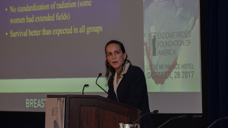
But, about 5 or 6 years ago, the Z11 trial came around. Basically, what they were trying to do, they were trying to separate out women that could potentially avoid an axillary dissection even if the sentinel node was positive. If we're talking about 5 and 10 years ago, when a woman had a positive sentinel node, we would go on to do the axillary dissection. This study was looking at women that underwent breast conservation, so it did not include women that had mastectomies, but it was looking at women that were undergoing breast conservation. They separated out the women, and one group of women had an axillary dissection if they had a positive node, and the second group did not have an axillary dissection. What they found was that there was no survival difference, and there was no difference in local recurrence.
Now, there were some problems with this trial. It failed to meet accrual. Women in the trial had knowledge of their disease in the sentinel node. If a woman had a node that was replaced with tumor, she was more likely not to join the trial. The women with worse disease probably were not included in the trial. There's also no standardization for the radiation that these women received. If a woman had a positive node, instead of just radiating the breast, the radiation oncologist would often extend the field and radiate the lymph nodes under the arm. It's not a perfect study, but, basically, it has with time proven that it's probably applicable to patients in today's setting.
We use this criteria in women that have tumors that are less than 5 centimeters, less than 3 positive nodes, and the patient does need to have radiation because the radiation probably is treating some of the lymph nodes under the arm. The concept is basically that, if a woman has a positive node, she's going to get radiation if she's having breast conservation and she's going to most likely get chemotherapy. The radiation and the chemotherapy probably sterilize any other lymph nodes under the arm that have disease. Therefore, we can spare these women axillary dissections. The risk of lymphedema long-term is going to be much less and so, really, overall, a big improvement in what we do from a breast surgery standpoint.
One of the other advances which has really come about more recently is this device, and this is the BioZorb. Basically, this device has clips within it, and then this is made of suture material. We place this in the breast, and I think I have a video. Let me go forward. We place it in the breast, and then we rotate the tissue over the device. This helps maintain the shape of the breast. Now, it's not replacing the volume, but it prevents the process where, if you have a big hole here, the skin will get tethered down, and you get a distorted breast.
Even without the use of BioZorb, you can use oncoplastic techniques where you rotate the patient's own breast tissue into the defect. You basically separate the tissue from the skin, you rotate the tissue forward, and you can prevent the pulling in that occurs with breast conservation surgery. This, however, also has the benefit of marking the area for the radiation oncologist, so it gives some support to the breast, it prevents the quick collapse of the tissue and the skin, and it marks the area where the radiation oncologist will give a boost of radiation. When a patient is getting radiation, they get the standard whole breast radiation, but there is a boost for about a week to the tumor bed. This clearly delineates where that tumor bed is, so it helps with the delivery of radiation as well so improved outcomes probably long-term. ... Basically, that's what I just said.
Just briefly, you're going to have a talk on this later, I'm going to mention there are improvements with breast construction. We are still doing implant reconstruction, but we are often using a woman's own tissue to create a breast. We remove the breast tissue, and we have to replace that space with something else. It's either an implant in women that don't have any extra tissue anywhere or it's filling that space with tissue most often from the abdomen but often as well, if they have heavier thighs, we can take tissue from the thighs and the buttocks. That's a free-flap reconstruction. You're going to hear a lot about that later.
I'm just going to give you a few pictures. This is a tissue expander. A patient undergoes a mastectomy, removal of the breast, and, in this case, obviously, the nipple and the areola. We put a spacer in at the time of surgery. The patient goes in every 2 to 3 weeks and has fluid injected into that space or tissue expander, and there's a second stage surgery where that expander is taken out and the permanent implant is put in.
This is a patient that had implant surgery. In clothes, you can tell that she's had the surgery. Outside of clothes, it's going to look pretty normal. You can see the scars here.
Now, this is a free flap, and this is a big benefit. I definitely have a bias towards using natural tissue if you can. It's not possible in all women. But, basically, if a woman has some extra abdominal tissue, the benefit is she gets a tummy tuck. Yes, there's a big scar, but basically she gets a flat abdomen. We take this tissue, and we fill the envelope that's created at the time of surgery with the tissue from the belly, and you get an excellent cosmetic result.
We try to do techniques where the scars are hidden, so these women, honestly, if you looked at them, we have women coming back to us and telling us that their doctors didn't understand why they didn't have their surgery, and the doctors are very surprised when they're told that the surgery was completed. This not only looks like a natural breast; it ages like a natural breast. To the outside, it feels like a natural breast. Really, we've had great strides made with breast reconstruction, and you're going to hear about that later in the day as well.
In concluding, breast cancer in young women is very challenging. There are very unique issues that arise with these young women. Fertility issues are certainly very important, and we still need to work on improving what we can offer these women, but there have been many technical advances that have improved not only the outcomes but the cosmetic results as well. Thank you.