Endometriosis Foundation of America 2014
Adenomyosis: diagnosis, treatment and impact on pain and fertility
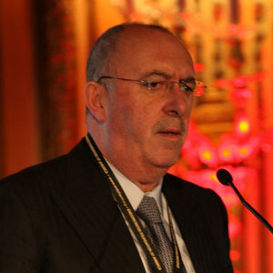
- António Setúbal, MD
Thank you Harry, thank you Lilo and thank you Tamer for the invitation to be here to talk.
There is really no controversies to talk about with adenomyosis but we have a lot of questions and that is what I am trying to bring here today. When you talk about adenomyosis it is a bumpy and endless road that nobody wants to take. It is quite a difficult disease. In order to make a simple definition you see that clearly everybody knows that adenomyosis refers to a disorder in which endometrial glands and stroma are present within the uterine muscle. So it means it could basically be diffuse and sometimes, but rarely, could be focal disease and not diffuse disease. Basically, we are going to discuss this a little bit.
As you know, for the definition ultimately there is an anatomopathology diagnosis but the question is, is it? Well, let us again discuss this a little bit and the answer is no. The incidence is not well known and it probably affects about 20 percent of women. In one histopathological study the incidence was 65 percent. Can you imagine? I have to stress at this point that I think it is important to know that the disease is becoming more aggressive and is affecting more patients and more young patients. That is the most important thing concerning young patients with adenomyosis.
The incidence just to bring you a couple of data in a study of 227 women with and without endometriosis have found that pelvic endometriosis is strongly associated with the thickness of the junctional zone, as everybody knows. Also the rate of increase is steeper after the age of 34. The junctional zone was thicker even in young women with pelvic endometriosis. In one study they found that women with endometriosis will have at least 90 percent of adenomyosis also. The pathogenesis is still unclear, which means not known but there are two major theories as you know. One is related to the endometrial invagination into the wall, and the second is the “De Novo” formation from Mullerian Rests.
As I said before we can see that in terms of types there is diffuse. That could be sclerotic or could be glandular or cystic, which means this is a type of adenomyosis that you can find mainly in young patients. Rarely are you going to find a very confined area where you have an adenomyoma. But usually it is also associated with diffuse adenomyosis as you can see in this specimen here. Again, glands and stroma in the middle of the myometrium, as you can see this is clearly adenomyosis.
When we talk about clinical manifestations we have to think that there is always progressive, or almost always progressive heavy menstrual bleeding. There is progressive dysmenorrhea, there should be anemia at the end. There is sub-fertility or infertility, dyschezia, urinary symptoms, pelvic organ pain and complications of pregnancy that we will talk about a little bit more in a couple of minutes.
Is adenomyosis related to sub-fertility or infertility – definitely yes. Why? Well we have a couple of reasons. When you have diffuse endometriosis all the myometrium is increasing so at some point it will occlude part of the tubes related to the wall of the interstitial part of the tubes on the myometrium. Then you have to think about the alteration in the ectopic endometrium that is dysfunctional or disruption on the junctional zone with inflammation, implantation, neovascularization and difficulty related to the embryo zone. There is…contraction of the uterus. Up to 50 percent of women with adenomyosis will present with dysmenorrhea and menorrhagia. That is a defective deep placentation and that was proved already by Campo a long time ago and by Stephan Gordts at the beginning of 2013. There is still a long way in order to go into…that I showed in the beginning. This means that this is very anal, still very anal disease.
When you look at this publication of this past year in February from the Japanese they are doing a lot of work in adenomyosis. When you look at the annual report of the Reproductive Endocrinology Committee from the Japan Society of OB/GYN they looked to all these details and what they figured out was something that was already mentioned in 2004 by Minegishi. As you can see there is sub-fertility no treatment established yet. There is up to 50 percent miscarriage, there is pre-term birth of at least 24.4 percent and the fetal growth retardation of almost 12 percent. There is obstetrical bleeding, preeclampsia is more when you have adenomyosis with a diffuse type and also intra uterine infections when you have the same diffuse type.
When you look at another paper concerning adenomyosis and sub-fertility, this paper was done by Tomassetti, it tried to obtain answers for question about adenomyosis. It was done in baboons and based on limited available evidence to support a causal association between adenomyosis and sub-fertility. Adenomyosis is associated with life-long infertility in baboons and is associated with impaired reproductive outcome after assisted reproductive techniques.
If you jump into another publication, this one concerning adenomyosis reduced pregnancy rates in infertile women undergoing IVF, if you look at the numbers you can see that there are 256 women without, apparently without, adenomyosis and 19 women with adenomyosis. Where you will find it is in the clinical and ongoing pregnancy rates which are completely different, 22 percent versus 47 percent in the group that has adenomyosis or the group that does not have adenomyosis. Actually you can also see that miscarriage was higher in human women with adenomyosis compared to those without adenomyosis. This means a huge difference between 50 percent and 2.8 percent.
We are in the presence of a not very clarified disease but we are starting to have some data about it. The diagnosis of histopathology is the gold standard but the question is can we not have the histopathology, why? The reason is because women want to get pregnant and they already have adenomyosis so you are not going to do a hysterectomy because you need to preserve fertility.
We have some other tools that are becoming more and more interesting, Consider that we have MRI, which means that you can…into for sensitivity around 88 percent and…specificity of 93 percent. You can see all the glands on the myometrium, the thickness of the junctional zone and asymmetric walls. But also, ultrasound in good hands can give you a good approach to adenomyosis. You can see the glands on the myometrium and again the thickness of the junctional zone and asymmetric walls. Again, sensitivity of 83 percent and specificity up to 85 percent.
Then you have historiography and hysteroscopy with the suspicion of bluish spots, the glands, the aspect of the strawberry lesion. It is not on the market yet and it has not been approved even in CE and far from that with the FDA, but Stephan Gordts’ biopsy again could be good help in the future. I will show you in a couple of seconds, laparoscopy and of course cystoscopy if that is the case.
So, MRI – you can see here clearly a big diffused adenomyosis with adenomyoma and that I went into difficult surgery. I do not have the time to present to you the end because I have to keep on time and we are delayed already almost one hour. After the control was there to still have diffused adenomyosis but at the end there is no huge… This patient wants to get pregnant and was sent by referral to me by the IVF people.
Again, more aspect of diffuse adenomyosis. As you can see here you can clearly see the difference between adenomyoma and myoma as I have shown you before. Here, this is a complete diffused adenomyosis and with cystic implants over there, asymmetric walls of the uterus. This is utero cells, 3D and again you can see that in good hands and with good experience some of the disease that you can find over there. This is a sclerotic form of adenomyosis, like this one too and this one. I stress the point of in good hands you also can see that is very classical for adenomyosis, which is the classical pearly state due to…when you do the fluxometry of the uterus, then again, the same.
This is a myoma and this is an adenomyoma in ultrasound. So again I stress the point that in good hands for some ultrasonography you can have a good diagnosis of the situation.
What about this? You can clearly see that this is absolutely a very bad case of adenomyosis in this hysterosalpingogram, the strawberry aspect in hysteroscopy and the bluish spots in hysteroscopy. Just to show you, because Stephan Gordts gave it to me just to show you that mainly in the future we are going to have the possibility of another good pathology report using this type of device developed by Stephan Gordts in Leuven. But again I stress the point that it is not approved, not even in CE. Actually it was approved in CE for breast cancer biopsies but not yet for the reason that it was blocked for adenomyosis and then you have laparoscopy. For those of us used to doing laparoscopy clearly this picture is adenomyosis, adenomyosis again and subsequently again you can see bluish coming up.
So treatment: well, for asymptomatic patients, expectation and no treatment. For the rest, well you have a huge number of drugs that were talked about because of endometriosis; we just talk about them in the morning so I am not going to go into details. But starting at analgesia and going down to the new SPRMS maybe with the new Ulipristal or the new type of Mirena, which is called Jaydess. I show it to you here. I do not know if it is approved in the United States yet.
For non-surgical treatment you have the uterine artery embolization and the so-called MRI FCUS ultrasound, but again, poor data and poor results. For surgical treatment of course – plus or minus BSO in the diffuse/focal, it is the gold standard. But for a few rare cases the focal adenomyomectomy as I showed you before in that picture – I do not have the time or energy to show you the film – it is very good in very selected cases.
The other surgical treatment, a cone resection with a Strassman like on resection. Then something that was already a question this morning about the double/triple flap partial hysterectomy developed by the Japanese school, I will go into that because there was a question this morning. And of course, ligation uterine artery ovarian – that is the same, there are few cases reported and the results are very doubtful. It is kind of what we could call a desperate surgery with no good data in order to see if any kind of those surgical treatments should be shown.
This is as I said before the Japanese technique of double/triple flap and this is the situation after ten years. As far as can be seen from Osada’s people they treated about 100 patients. And from what I can see from here 16 became pregnant and 14 went to the end. The symptoms recurred in only four cases out of the 100 for surgery. But, if you jump into the details of this operation, this is an open operation. This is not laparoscopy it is laparotomy. It is a very hard operation and I am not going to enter into the key points of the situation of how to do it. I think I have just to give you the idea – of course you are going to lose one of the tubes. At the end we see the aspect of the uterus according to the double/triple flap technique.
To save time I have skipped the film. I thank you for your attention.
Dr. Liselotte Mettler: Can you come up, the two speakers?
Dr. Harry Reich: We are sort of pushed for time so we are limited to three questions from the audience. But this is what endometriosis surgery is all about and I think you saw really good examples, great examples, of two of the more popular techniques. I emphasize these, what we saw, were stage 4 cases and are not probably the every day for many surgeons. But, of course, for somebody like Tamer, for our faculty, this is probably what they see on a regular basis. Questions?
Dr. Liselotte Mettler: If there are no questions from the audience I would like to ask Dr. Advincula what are the expectations for the future of robotic surgery with smaller devices, cheaper devices, technically affordable robotic three dimensional instruments?
Dr. Arne Advincula: I suspect that, well I have seen some of the future prototypes and it is probably getting smaller, I do not know how quickly things will get cheaper. I think there is a point of diminishing return in terms of size, at least speaking as a gynecologist because when you are dealing with very densely fibrotic tissue that is just welded to the pelvis, or you are dealing with large myomatous uterus for example, there comes a point where you do not want too small and too delicate an instrument. Otherwise it is really not going to be useful for you so I think there is a threshold that you eventually cross where you are really not making any forward progress. I think miniaturization will happen. I think if it does it will find its home better in other specialties like otolaryngology for example where they are using robotics now but they clearly will benefit from miniaturization of some of the instrumentation. But, certainly for some of the work that we do, or at least I do, I do not want it to get so small that I will not be able to get done the job that needs to get done. That is really where I see things.
Dr. Tamer Seckin: (Cannot hear question/comments – without mic.)
Dr. Jon Einarsson: I think that if we are honest with ourselves I think that most of the time we leave some disease behind. It does not matter who is operating. In principle, because some of this disease is microscopic we cannot see it and I think also that obviously we do our best to get all of the fibrotic tissue out of there. But nobody really knows very well how that affects the patients because unfortunately these patients are complex; they have multifactorial cause for their pain. In order to figure out these questions we need a very large sample size of patients because there is a lot of noise in that data set.
But I strive to remove all of the endometriosis that I can see but I think that if we are honest with ourselves we are leaving some of it behind.
Dr. Arnold Advincula: I agree 100 percent with Jon. I think that that is an important part of being a surgeon. I actually think that the hardest part about being a good surgeon is knowing when not to do something and not to go too far and be too ultra radical that you actually trade one issue for another and potentially compromise the patient’s function. Yes, I agree with Jon, it is hard to know you have gotten everything and I have not really found a technology that I love that says it is going to do it all. But even if I did have that technology I think that that is the art of medicine knowing how far to push technology without compromising function.
Dr. António Setúbal: Just a question about when Tamer said about irrigation or suction. Jon and I operate almost the same way because we belong to the…school and that style so let us see that you lose the nodule on the rectum we try to put another into the vagina wall. But the main thing in order not to lose the cleavage plane is do not irrigate. If you irrigate you are going to lose all the cleavage plane before you get the lesion completely…
Dr. Harry Reich: Let me just make a comment. I think we talk too much about microscopic disease. As surgeons typically we can examine the patient before the surgery. We can find out where she hurts and our surgery should be directed to the areas where she hurts to remove those areas specifically. Most of the time you can feel them if you do a rectovaginal exam in the office but many gynecologists do not do rectal exams I know that. It should be part and parcel of any endometriosis surgery to find out where they hurt. If you remove those areas, especially endometriosis glands, stroma and fibro-musculature tissues surrounding the glands and stroma – that has probably been there for the last ten to 20 years, and if you remove it all it will take a long time with active hormonal stimulation for it to re-accumulate. They should be pain free.
Dr. Liselotte Mettler: We have a question from the audience.
Audience Member: (Cannot hear question/comments – without mic.) …have better visualization. I came across a paper recently on microscopic disease and by traditional laparoscopy the rate of microscopic disease with well defined, good definition of…tissue and with…proximity to…the rate of microscopic disease is found to be negligible. So I guess my question is how can you improve on that..?
Dr. Arnold Advincula: I did not say that in my talk. In my talk the point I was trying to make was that there was a lot of media emphasis and marketing emphasis when I put that slide up that they are saying that the robot allows you to see better, therefore you can cut it out better. My point is that the robot does not necessarily guarantee you are going to neither see anything better nor cut it out any better. My point there is that you have to know the biology of the disease. You have to have recognition as the surgeon. No robot will compensate for that. Even some of these fancy things like the indigo sign in green florescence imaging – I am not buying into that because I have seen enough people that have come through my office where they supposedly had that done, had a robotic procedure done in fact by places here in the city and you go and do their exam and there is a big nodule that somebody missed just by not doing a rectovaginal exam. They are relying too much on something to light something up that was clearly, obviously there.
I think that that is the point I was trying to make. It is really intrinsic that the surgeon knows what they are doing and then you could really make that technology sing for you. Otherwise it is dead in the water.
Dr. Harry Reich: We operate on fibrosis really. You do not see endometriosis glands and stroma on benign looking peritoneum. Inside the fibrosis that is where you find the glands and stroma.
Dr. Liselotte Mettler: The basic examination of the gynecologist is still the rectovaginal exam. So many gynecologists do not want to do that anymore because it is painful for the patient. It is not a nice procedure but it is as basically important as the ultrasound is.
Dr. Arne Advincula: If not more.
Dr. Harry Reich: One last question?
Audience Member: Hi Arne, welcome to…good to know that you are nearby. I will send you our patients. I wonder Jon…if some of the studies that are looking at the benefit of robotic versus traditional laparoscopic surgery involve regular surgeons like me? I am a good surgeon. You are a fantastic surgeon; I wish I could do what you do. But I do get to work on a hell of a lot of people. The robot system has absolutely transformed gynecology in my community. I am in a group of eight physicians. We have young surgeons, older surgeons, do lots of vaginal hysterectomy. For years we just could not really significantly decrease our abdominal hysterectomy. We were running at about 40 percent. In the course of three years we have our hysterectomy rate down to three percent. That is a lot of people that have been positively affected by that technology.
I appreciate the studies done in tertiary care, specialized institutions but are there studies that are looking at regular people and the societal impact that this technology can give?
Dr. John Einarsson: I think that is a good question. I think the enabling part the most enabling part of the robot is you get surgical volume. A lot of times you get sort of a center of excellence or people that are doing a lot of surgeries. They can sort of act as a fertile ground for teaching the rest of the staff. The same thing that you just described has happened across the country in similar hospitals with regular laparoscopy too.
What is happening with this is that with our mixed fellowship we are producing people who are competent laparoscopic surgeons and then they are going into the community. They are acting as leaders or mentors for their staff at their hospital. I can say that I was one of them. I came to Brigham in 2007. At that time they were doing 70 percent abdominal hysterectomies. We have a staff of 40 surgeons and a volume of 1100 hysterectomies a year. Now we are down to less than 10 percent. And it is not just my doing but I think I was instrumental in getting that ball moving and mentoring a lot of people that are now doing it on their own. We do it all straight stick. I do not think it is the robot per se because I think there is a little bit more moving parts for you to learn to do the robot. But it is enabling you to get patients and again have more volume and then improve your skills. I am not sure it is the robot itself but I hear that argument a lot.
Dr. Harry Reich: I think we went to sort of a different tangent on hysterectomy instead of endometriosis. The company will certainly try to promote as much as they can with the robotics.
With that I will leave it to our next session. Thank you very much for your attention. Let us take a stretch and we will try to get started within the next five minutes? Is that all right?


