As you know last year Linda Griffith was our keynote speaker and honoree for her work, but this year our keynote speaker, science section, is a very well known writer and stem-cell researcher. Her writings have been an inspiration for me I must say. She came all the way from Monash University, I would like to introduce to you Dr. Caroline Gargett.
Caroline Gargett, MD, PhD
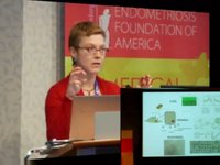
Well, firstly while it is just loading up, thank you very much Tamer and the foundation for inviting me to come and speak to you. It is indeed a pleasure, and I have enjoyed the conference so far.
I am going to talk to you a little bit today about endometrial stem progenitor cells and I have broadened out the talk to cover their role in a number of different areas in gynecology. Firstly I would like to just cover a little bit about endometrial regeneration, the stem cell hypothesis and gynecological disease. Then give you some of the evidence that we have generated to tell us that there are endometrial stem progenitor cells in human endometrium, and markers and ways in which we are able to identify these cells, and then to look at where we are using this knowledge now in gynecology. I am actually going to show you a very early diather on endometriosis. We are looking to do a tissue engineering application and we have got some evidence too for cancer stem cells in endometrial cancer.
One thing about endometrium it is a highly regenerative tissue as many of the audience would already know. During the menstrual cycle this functional layer of endometrium grows. It is about a centimeter worth of mucosal tissue growing in a very short space of time, not only does it regenerate in the menstrual cycle, but it regenerates following parturition when the endometrium is resected, at times it regenerates. And post-menopausal women who have a very thin endotrophic endometrium, if they are placed on estrogen only hormone replacement therapy you can grow their endometrium like a thick cycling women’s endometrium and it is able to support pregnancy to full-term. So this endometrial tissue is very regenerative and because of that we, and others, hypothesized that there would be a stem cell population, particularly we thought there would be in this basalis layer, which remains, which is like a germinal compartment and it is not shed during menstruation. We thought there would be two types; we thought there would be an epithelial type and we thought there would be a stromal type because there is a lot of stromal tissue supporting those endometrial glands.
The reason we really were interested to find adult stem cells in endometrium is that there are a number of gynecological diseases which are associated with abnormal endometrial proliferation, which we thought could really be considered an endometrial stem cell disorder. Endometrial cancer is the one that comes to mind first. Here we would think that there is a mutated stem or progenitor cell that is responsible for say initiating that tumor and for many of the other features of tumor growth, and metastasis and recurrence. But in endometriosis here we would think that perhaps a normal stem progenitor cell is shed by retrograde menstruation into the peritoneal cavity to establish an ectopic implant. Also, in adenomyosis, which is ectopic growth of the endometrium in the muscle layer, we think that perhaps here again there is a normal stem progenitor cell an abnormal niche regulating that stem progenitor cell that is responsible for the features of adenomyosis.
Then we could look at the other side of the coin where there is perhaps a lack of, or ineffective stem progenitor cells and Asherman’s syndrome and even when you ablate the endometrium really there must be damage or loss to the stem progenitor cell population. There is a condition in IVF where the endometrium is very thin and no matter what hormonal support is provided the endometrium just will not grow thick enough to support implantation. Here we think there may be a diminished activity of normal stem progenitor cells.
I just want to give you a little bit of background on adult stem cell biology, so I hope you understand the rest of the talk. In any tissue really there is a hierarchy of cells, with the stem cell at the apex, the master cell and it is the rarest cell. At the bottom are the worker cells or the differentiated functional cells of the tissue. There are three properties that set the stem cell apart from the rest of the tissue cells; they are the fact that it can self-renew or replace itself and keep that small pool of stem progenitor cells, but also their ability to differentiate at the same time to produce the functional differentiated cells, and they also have a high proliferative potential. Just a single stem cell division can produce a large number of cells and the proliferation of the stem cell is really amplified by these intermediary cells, which we call trans-amplifying cells. Paradoxically though the stem cells do not proliferate very often at all. In fact, really we regard them as a quiescent cell. It is these cells here that do all the proliferation. In vitro we can actually set up that little hierarchy by cloning the cells or seeding them at a very, very low density in culture so that single cells set up a little growth of cells, and they are called colony forming units.
When we started to look in endometrium, which had never been examined in this way before, we had no markers and no way of knowing whether there were stem progenitor cells there, we went back to first principles and borrowed from the hematopoietic system and we did these colony forming unit assays. We took hysterectomy tissue because we needed to have the basalis, we dissociated to single cells and we separated the epithelial and stromal cells and we just seeded it at very low density on these culture plates. You can see little clones of cells are being produced. You can see the frequency of them as well. In particular we were interested in these large clones, which were quite rare that we thought might be initiated by the stem progenitor cell. We found, importantly, that these clonogenic cells were present in inactive endometrium, not post-menopausal endometrium. We did not really find any particular difference in their frequency, whatever stage of the cycle we looked at. Then we took those large clones and we looked at those three cardinal properties that I told you about before. We looked at self-renewal setting ourselves at incredibly low densities and we just re-cloned those cells after we could harvest individual clones and re-clone them. This ability to re-clone tells you that there is self-renewing cell layer that can initiate another colony. We were able to do this for the epithelial and stromal cells three to four times indicating quite extensive self-renewal capacity. We also looked at their proliferative potential. Here would take one of those clones and we would culture them out till they could not culture anymore, and the epithelial and the stromal seed a few produced about single cell production of about 30 to 34 population doublings. This is the production of several billion cells from a single cell.
Then we looked at differentiation. For the epithelial clones we took the secondary clones, we cultured them in a three dimensional medium and we were able to produce really quite large gland-like structures. When we took the stromal clone, we took the secondary one, and remember this started from a single cell originally in the primary clone, when we grew them in specific induction media we were able to differentiate them down to smooth muscle, to bone lineages, to fat lineages and to cartilage lineages. This tells us that these cells are multi-potent and these mesodermal lineages indicate that there are mesenchymal stem-like cells. This data tells us that in human endometrium there is an epithelial progenitor cell and a mesenchymal stem cell.
Now those assays are all done in vitro and the gold standard for stem cell activity is to be able to reconstitute the tissue in vivo, in a living animal from one of those single cells. That has not been done yet but what has been done by a group in Japan they have been able to transplant just singly dispersed endometrial cells, a mixture of epithelial and stromal cells under the kidney capsule, and they have grown endometrium that looks and functions just like native endometrium in the woman, and it responds to hormones. But even this assay, this in vivo assay and in vitro assays they are all retrospective and there is no way that we can recognize what that cell is that is initiating and showing these adult stem cell properties. We have just shown stem cell activity. We have been very keen to find markers that will be able to isolate these cells so that we can study them. We could look at them and some of the types of technologies you have already heard about this morning and also to find out where they are and investigate their role in gynecological disease. We have been looking for markers for both those cell types.
Work with the epithelial line has been really incredibly difficult. This is really quite challenging work. We have really done a screening, searching a panel of markers on endometrial cell suspensions, in epithelial cell suspensions we do it by flow cytometry. We then look at immunohistochemistry and we are looking for expression of those markers in the basalis, it may be stronger in the basalis epithelium than in the functionalis. We are looking for heterogeneous staining because we know that not all the cells will be a stem progenitor cell, they are quite rare. This particular one, this H3D12 is one that we have investigated further and in fact this sort of summarizes those 30 antibodies that we have looked at and here is this H3D12 which was quite specific for epithelium and exhibited some of those distributed by immunohistochemistry in the endometrium. We then devised complex flow cytometry, in fact sorting protocols, to examine the function then of those markers, if we separated the cells according to that marker expression. We have done excluding a lot of cells but using EpCAM and the H3 marker we can see that there is a number of populations there. Each dot represents a cell and we were able to sort those cells, so here we have sorted H3D12 negative line because it is negative on the bottom axis here but expresses EpCAM and we rarely got any clones. But my student was able to do a little bit of culture. When we cultured those double positives cells that express H3 and EpCAM we got lots of clones. When we cultured or did the serial cloning of the H3 positive cells but EpCAM negative we got some quite large clones which we are very excited about. And this data just shows, it is still preliminary data, and just shows that data it is color coded to help you follow, but this is this population here where we got more cloning than we did in the other two populations.
But when we applied the more stringent test, the self-renewal assay, that serial cloning, and this is just showing our preliminary data here, this is the primary, the secondary, the tertiary clones and you can see that the green ones, the double positive cells, five out of six patients we could clone in primary culture and we got tertiary clones out of the two out of two that we have tried so far. This is work in progress. For the purple ones, for the H3 positive outcome negative, they could get secondary clones in half the samples, but no tertiary clones in the two samples we have looked at. So we are more interested in this double positive population as a result of this assay. But what we can see here when we look at both those populations compared to the large cells which we have put through the sorter, which damages the cells quite considerably, we still have enriched further cloning and self-renewing cells in these populations compared to our live population.
When we have looked at the stromal cells we have been far more successful and it has been a lot easier because they are easier cells to work with and we can borrow from other systems because mesenchymal stem cells have been looked at elsewhere. We put together two markers, the CD146 and the PDGF receptor beta and you can see this small population here and we have demonstrated that these contain a lot of the clonogenic stromal cells in human endometrium. It gives an eightfold purification over unsorted cells and about a 17-fold over other populations there. We have shown that they are multi-potent, they show typical mesenchymal stem cells surface signature and when we looked at where they were, so when we did confocal microscopy to look at their co-localization of those two markers we found, it is probably hard to see on this image here, but the yellow cells, the co-localizing ones, are right next to the endothelial cells around blood vessels. So they are perivascular cells and we think they are pericytes and further work in other systems has corroborated that it does appear that mesenchymal stem cells are a pericyte-type cell. When we looked in endometrium to see where these perivascular cells were, this double positive CD146 and PDFG receptive beta, we found that they were indeed around blood vessels in the basalis layer but we also found that they were in the functional layer. This is quite an important finding because this tells us that these endometrium mesenchymal stem cells will be shared in menstrual blood. This is quite important in the concept of endometriosis.
However two markers are not that easy to deal with if you have to set up complex facts and flow-cytometry and really we are looking to use these cells in tissue engineering for example, and I will just briefly mention that. So a single marker is far superior to work with. We have also then screened out stromal populations for a number of perivascular markers. These are still yet unknown markers, there is a W5C5 and you can see an endometrium it is around blood vessels. There is a small population. When we cloned off those populations virtually all the cloning activity is in the W5C5 positive population. You can see that here. We have undertaken our self-renewal assays they differentiate and importantly with these we have been able to transplant them under the kidney capsule and generate human stromal tissue.
This particular marker now is really important for us and we want to use this for a number of applications and I am going to show you now how we use this. The first item I will talk about is endometriosis and this of course is the focus of this meeting. Our hypothesis is that endometrial stem progenitor cells may be shed in the retrograde menstruation and gain access to the peritoneal cavity where they will establish an ectopic implant in susceptible women. This is the first time I have presented this data and it is really preliminary but I thought at this forum you would be interested to see what we are trying to do now that we can look at some of these markers. We have been working with a gynecologist, Gareth Western. When he is doing laparoscopic surgery on women who are on day two of their menstrual period, we are obtaining the peritoneal fluid, the shedding endometrium and peripheral blood. Our controls are women having a tubal ligation. If we look at the menstrual blood first, endometriosis and control, we find clonogenic cells in all of them, whether they have endometriosis or not, which corroborates what I was saying before. When we look at the H3D12 cells, we have only done three samples of each and we cannot really see any difference at this point in time in menstrual blood. But when you look at the W5C5 cells it is not significant at this point because we have not done enough samples, but there is a trend and it does look like there is an increased number of the W5C5 positive cells in the menstrual blood.
When we look at the peritoneal fluid we found five out of seven patients we have looked at have produced endometrial cell clones but we have also found in about half of our control ones the same thing. It is hard to know whether it is really quite specific and it is actually opening up different ideas as to just how endometriosis may occur. But I would have to say that we have done three severe cases and each of those have been the only time we have seen epithelial clones in the peritoneal fluid. When we have looked at the H3D12 cells we do not really see a difference, but for the W5C5 cells again there is a trend. I think we need more samples to be certain that there may be increased numbers of these in the peritoneal fluid of women with endometriosis.
Now, when we look at peripheral blood, because let us face it menstrual fluid is predominantly peripheral blood, we have not found any clones, any H3D12 cells or W5C5 cells so they are definitely coming from the endometrium. The other thing that we are focusing on, using the single marker that we were able to isolate, the mesenchymal stem cell, and because mesenchymal stem cells really have quite a lot of properties that make them ideal for tissue engineering purposes, we are looking at a condition called pelvic organ prolapse, which I know the gynecologists in the audience will know, but this really is a very common problem that affects women as a result of child birth injury where the bladder and the bowel and the uterus can prolapse into the vagina. This is just showing a cystocele, or a severe bladder prolapse. The symptoms are incontinence and bowel incontinence and sexual dysfunction. It really is a major clinical unmet need because treatment is reconstructive surgery and it affects very large numbers of women.
But the surgery has not really been as successful as the urogynecologists would like. We are proposing to use our endometrial mesenchymal stem cells in an autologous manner for treatment of pelvic organ prolapse to really improve the outcomes of the reconstructive surgery. We are proposing to use endometrial biopsy to obtain the cells because we know that they are in the functionalis layer where we have methods now for isolating them, this shows a two marker method but we use W5C5. We have been developing an optimizing culture condition of these for clinical grade, a good manufacturing practice which is what we need to do. We have been scaling up the culture because we need to have large numbers of cells and we are doing this in three dimensional on beads. We are developing new mesh materials of scaffold materials that will carry the cells into the tissue that is being reconstructed because the current materials that are available now have been designed for abdominal hernia. And really the vaginal wall is a far more elastic type of tissue and requires really quite a different type of material. We are working with C SIRO, which is a big, main research organization in Australia and they have been working with us, the engineers to devise new scaffolds. We have based it a lot on the mechanical properties of human vaginal tissue itself and this just shows us testing it. We have picked our three materials, the polypropylene is what is in current use. What this graph shows is that polypropylene is very, very strong, much stronger than human vagina, which is here in red. We have chosen three materials that are about as strong as human vaginal tissue but across this XX is far more distensible, able to perhaps stretch and return to where it was than say polypropylene.
What we are doing is we want to combine these novel synthetic scaffolds, which we have had to add gelatin fibers to with our endometrial mesenchymal stem cells to produce a tissue engineering construct. We found that our cells do grow on the two materials that we are currently developing, and they are the green cells there. We found that the pores are very, very large and because of that we cannot really deliver enough cells, so we need to fill those pores with something that our cells can grow on. We are using a gelatin collagen at the moment and you can see our cells growing in between these fibers. Then we are testing them. At this stage we are only just testing the materials that we have designed in a rat abdominal hernia model. This just shows some of our earliest explants from just transplanting or implanting the mesh and we are doing a lot of biocompatibility testing, mechanical properties, histologies, just shows the area, which is really ongoing work. And yes, we will eventually take it up to much larger animal models cells and our goal is to translate this into the clinic for women with pelvic organ prolapse.
Finally, I want to talk about cancer stem cells, and again I want to take you back to that hierarchy in normal tissue. Just like that in cancer tissue there is a hierarchy as well. There is evidence from leukemia research that the cancer stem cell could be derived, or can be derived, from the stem cell itself, the normal one, which undergoes mutations, say in the tumor suppressor genes for example. But equally it has been shown that much more mature cells in a tissue, such as the progenitor the trans-amplifying cells, can undergo mutations say in the self-renewal genes that may make them constitutively active and produce what we call this cancer stem cell. This cell occasionally divides and produces a hierarchy of cells which is the tumor itself, which is quite a heterogeneous collection of cells in that tissue.
When we treat cancer with chemotherapy, chemotherapy and radiation therapy attack the dividing cells, and so you kill them off. But they do not really have much effect on these quiescent cancer stem cells so they remain. Then when we take the chemotherapy away the tumor re-grows and this is what causes recurrence and metastasis of the disease.
We have looked in endometrial cancer to see whether we could find a cancer stem cell in this tumor. We have indeed shown that there are clonogenic cells, we have shown there are tumor initiating cells and this just shows a snapshot of some of our work. This is the primary tumor. We have transplanted single cell suspensions in quite low numbers, this is just one. And we can produce the same tumor as the patient’s original tumor and it shows the same differentiation markers, estrogen and progesterone receptors for example as the original tumor. So we are indicating a differentiation in vivo and we have shown self-renewal. We can re-transplant those tumors and generate new tumors. We have also shown their express pluripotency and stem cell renewal genes.
And finally, just to summarize our work so far looking at endometrial stem progenitor cells in gynecological disease we have shown that human endometrium contains rare populations of an epithelial progenitor, which we may be able to sort out using that H3 marker. We have a mesenchymal stem cell population and we have two types of markers that we can use to isolate those cells. We know they are shed during menstruation. We think they will be involved in endometriosis lesion development because we found some of these cells in the peritoneal fluid. We have not looked at adenomyosis but we suspect that they are involved there, and also we think they may be involved in being able to generate a thick endometrium after biopsy. Recent studies have shown that in IVF patients that those who have thin unresponsive endometrium that if they biopsy those women even in the cycle before or during the same cycle of implantation that they double the rate of pregnancy. What I am thinking that might be happening here is that that injury, the actual currating or biopsying is actually damaging the endometrium. When we damage other systems, organs, the stem cells overproduce the mature cells like the bone marrow system for example. So, what I am thinking here is that this biopsying, even in the preceding cycle, activates the stem progenitor cells to produce a much thicker endometrium. We have also shown that there are cancer stem cells in endometrial cancer, and finally, we are looking at the potential use of endometrial stem progenitor cells in a cell-based therapy for pelvic organ prolapse.
And last, I would like to acknowledge my team, and there are many of them who have contributed to the work I have described today, and some of them are shown here in bold. Some of them are clinicians who I work with quite closely, and also the team at C SIRO, the engineers, and my German collaborator Hans-Jörg Bühring who has given us this panel of antibodies that has facilitated our work in a huge way.
Thank you for your time and attention.