Endofound Medical Conference 2017
"Breast, Ovary and Endometriosis"
October 28, 2017 - Lotte New York Palace Hotel
Mechanisms of Survival of Stem Cells in the Cancer Milieu
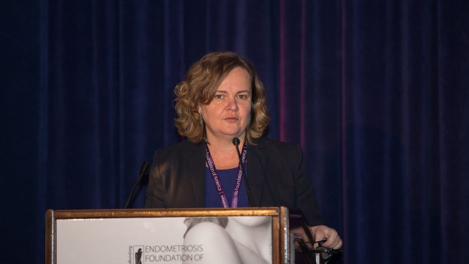
Daniela Matei, MD
Professor, Department of Northwestern Medicine Feinberg School of Medicine
Transdisciplinary conference. I have learned a lot this morning by medical oncologist, but my focus is on ovarian cancer. I'm a cancer biologist, so learning about endometriosis this morning is giving me a little bit of pelvic pain already.
So, the focus of my talk is mechanisms of survival, and perhaps new therapeutic options that could target cancer stem cells. These are different type of stem cells, not the embryonic stem cells, not the adult stem cells, but the stem cells that give rise to ovarian cancer.
And I will try to make an argument that perhaps the inflammatory environment present in endometriosis affect soil for this cancer stem cells to survive, and to perhaps proliferate and initiate tumors.
We have already had, earlier in the morning, that women with endometriosis have a higher risk of ovarian cancer and, very interestingly, the type of cancer that occurs in women with endometriosis is very distinct. Histologically, these are clear cell carcinomas, or endometrial carcinomas, or the low-grade series tumors.
Additionally, this type of endometriosis associated ovarian cancer, share unique molecular abnormalities, and particularly mutations in the ARID1A gene, which is a gene that's involved in regulation of transcription.
So, this have being said, a few questions, of course, arise. One is, "How should women with endometriosis be screened for ovarian cancer?" Or, "Do they have to be screened? Are there specific treatment modalities to remove endometriosis to prevent the risk of ovarian cancer? Is that necessary?" And one that's more interesting to me, on an intellectual level, "If this epidemiological risk is there, do we know how biologically endometriosis might be driving ovarian cancer? Is that even the case?" I would argue that that is the case, so we discuss regularly in our tumor boards, cases of patients that have endometriosis and associate it with ovarian cancer.
Here you can see a surgical specimen from a patient that had endometriosis. The area of the endometriosis under the microscope is illustrated here. Close by, you can see an area where inflammation is present. Next to it, an epithelial curse. Then, there's an area of borderline tumor associated with endometriosis. And then an area of real tumor.
So this phenomenon is real. And argues that there is a biological link between the two entities.
So what can be that connection? I presume you'll hear in the next talk about the potential genetic mechanisms that might may be common between precursors of endometriosis and precursors of cancer. Evidence for this comes from all the studies that have used a loss of heterozygosity analysis and found common LOH events shared between endometriosis and ovarian cancer.
But this here, we saw a very interesting report in the New England American Journal that documents, that presence of cancer associated mutations in very known oncogenic pathways, ARID1A, PIK3A, KRAS, that occur in benign [inaudible 00:04:46] endometriosis. Whether those [inaudible 00:04:48] of endometriosis will go on and become ovarian cancer remains speculative.
But then another mechanism non-genetic might be a fuel by the inflammatory microenvironment that is associated with endometriosis, which was very elegantly described this morning by Dr. Taylor. And indeed, the presence of a foreign endometrioid tissue in the peritoneal environment, causes recruitment and activation of macrophages. These, in turn, release cytokines on endogenics which, recruit endothelial cells, fueling blood vessels formation. And in this environment, cell proliferation is promoted, apoptosis is inhibited, and cellular invasion, again, is promoted.
The process of epithelial to mesenchymal transition, which is a fundamental hallmark of cancer, is a shared process between endometriosis and ovarian cancer. Not ovarian, any cancer. And basically means that transition of epithelial cells from this very nice cuboidal shape to an invasive, motile shape, this is favored, promoted by cytokines growth factors, reactive oxygen species, oxidative stress in the peritoneal environment.
And this CMT process is directly linked to stemness. So what are cancer stem cells?
Over the past 10 to 12 years, the concept of cancer stem cells has been advanced for solid tumors. These are rare, these are presumed to be rare on populations that may represent 0.5% to 2% of all cells within a heterogeneous tumor. These cells have the ability to self-renew, to generate more stem cells, but also to differentiate in daughter cells that have differentiated characteristics.
The stem cells are quite plastics. There has been recent reports that no-stem cells can be driven to become stem cells, and that's possibly one mechanism through which this can occur in endometriosis. They are generally quiescent and they are driven out of quiescence by certain stimuli. Many of them are not known, they may stay dormant for long periods of time and then reactivate. They have epithelial-mesenchymal phenotype, they are resistant to chemotherapy and radiation.
So, do ovarian cancer stem cells actually exist? The clinical course of ovarian cancer would suggest that these cells do exist. Because ovarian cancer is not, it is a fatal disease but it is not immediately fatal. In fact, most patients with ovarian cancer have fantastic responses to chemotherapy. 90% of them will enter a complete remission, and if they don't, I usually wonder if they actually have ovarian cancer, maybe they have a misdiagnosed colon cancer, because ovarian cancer is really remarkably sensitive to both chemotherapy and radiation.
However, these periods of remission, these are shown here as the CA125 number, it goes down from over a 100 after surgery and treatment, to normal levels it will stay down, but then, eventually, it will recur. And then, upon further treatment, this number goes back down, patient enters on other remission. But typically, the remissions are less complete and shorter, and they become shorter and shorter with treatment, until this disease becomes ultimately fatal.
So this is really consistent with phenotype driven by cancer stem cells, where by a tumor that's heterogeneous, it has a few stem cells and many other differentiative cells, when it's hit with treatment, chemotherapy or radiation, we get rid of the majority of tumor cells. However, this quiesce in stem cells, they persist and they stay quiescence for a while, until under the influence of some stimuli from outside, they wake up and they start proliferate and get rise to recurrent tumors. Many of these tumors then become resistant to treatment.
So if we could eradicate these cancer stem cells, perhaps there is a cure.
The first reports of ovarian cancer stem cells date back about 10 years, a group from Harvard defined phenotypes of cancers stem cells as these cells that are able to exclude the hoechst dye that are resistant to treatment. And when injected in mice, these cells called the side population, generate tumors as compared to the non-side population, which are not tumorigenic.
Subsequently, Dr. Nephew, in Bloomington, whom our group has collaborated for many years, identified CD44, CD117++ cells as cells that have these stem characteristics, self-renewal, tumorigenicity, chemo-resistance. And over the past 10 years, the number of publications looking at cancer stem cells has expanded, has increased, several markers have been proposed to identify these cells. The commonly used markers today are CD44, CD117++. These are really rare cells, they represent about 0.1%, 0.2% of tumor population.
And more recently, ALDH+, CD133+ cells also have tumor initiating capacity. These cells are easier to work with, they represent maybe 2% to 3% of cell population as I will show you.
Another concept that has been demonstrated by [inaudible 00:11:50] group is that cancer stem cells, when you grow them in non-radiant, non-differentiating conditions, they tend to form spheres. And we used these assay sphere formation in our subsequent experiment. I mentioned earlier that inflammation may drive tumor initiation. In 2008, Dr. Weinberg, at group at Harvard, demonstrated that breast cancer model, that non-cancer stem cells can be driven to become cancer stem cells by stimulation with TGF-beta, or by transduction of transcription factors that are stimulated by TGF-beta.
And we also show that in ovarian cancer model of that TGF-beta, which is abundantly secluded in the peritoneal environment, can stimulate cancer stem cells to form spheroids, both in cancer cell [inaudible 00:12:53]. As well as in tumor cells derived from patients.
Furthermore, IL6, which is a pro-inflammatory cytokine, stimulates or increases the number of cells with stem cell characteristics. This ALDH+ cells, when you treat them with IL6, the number of ALDH+ cells increases. And in fact, IL6 acts directly on the promoter of ALDH1 in using its expression and treatment with an inhibitor to IL6 blocks a ALDH expression.
Furthermore, knockdown of IL6 using genetic models significantly decreases the number of ALDH+ cells, as well as formation of these spheres, suggesting that TGF-beta, IL6, and perhaps other pro-inflammatory cytokines clearly drive the proliferation of stem cells, and might even transforms non-stem cells into stem cells.
So, we get now to the more immediate interest of our group. We are interested to find what are some of the vulnerabilities of these ovarian cancer stem cells. We referred earlier how important diet, and exercise, and metabolism is to the initiation of cancer. So, we ask the question, "Do cancer stem cells have unique metabolic characteristics that are different than non-cancer stem cells?" We hypothesize that may be the case, that's kind of an interesting thing to speculate. But how do you prove that?
The problem with the cancer stem cells is that they are very rare. So you cannot do the regular assays, mass spectrometry, et cetera, because you don't get enough cells. It's easy to speculate, but to really provide the evidence is a little bit harder.
So, we were fortunate that at a meeting, I met with a physicist, who developed a microscope using raman spectroscopy, and he said that he can image just a few cells. So the principle of this technology is that when you shine a laser light on the cell, the reflected waves are impacted by the composition of the cell. So, that you will get, eventually, a spectra of these peaks, that are taller or wider, which corresponds to the biochemical composition of the cell. It's quite complicated, I don't get it totally, but...
So, we set out to compare the cancer stem cells to the non-stem cells, and as I told you before, we selected the cells based on expression of ALDH and CD133. As you can see, it's about 3% or 2% of cells in cancer cell lines or in human tumors have these characteristics. So, when we did the first experiment, literally, it was just kind of an experiment, just let's see what we find. We observed that there was a clear difference, there were two differences: one at this wave length, which subsequently we decided it was noise. But we found a very reproducible difference at the 3,000 nanometer wave length, which apparently corresponds to a difference in saturated lipids.
So, of course we validated this in different cancer cell lines, as well as in cells from patients. And then, we also looked at cells growing as spheres versus monolayers, because the spheres are enriched stem cells, and we always got the same difference in this particular peak. We then went to confront these findings by using mass-spectrometry and we found that unsaturated fatty acids, where indeed increase in cancer stem cells, compared to non-stem cells. And then we ask, "Why would that be?" Well, the conversion of saturated fatty acids to non-saturated fatty acids is mediated by an enzyme SCD-1 or delta-9 fatty acids desaturase. So we measured this in the cancer stem cells, and found that it was increased in expression.
So we did subsequent experiment using inhibitors for this fatty acid desaturases, and perhaps our biochemical results don't matter as much, but what did we see with the cancer stem cells? When we use these inhibitors, we observe that proliferation of the spheres is completely blocked. This work was done by Salvatore Condello, a very talented scientist in my group. We found that the use of the inhibitors blocks expression of the other high dehydrogenase enzyme, as well as the expression of some of the stem cell factors.
We did similar experiments by using direct genetic down-regulation of SCD1, because some of the inhibitors might have off target effects, and we basically observed the same inhibition of the ALDH1, expression and of the transcription factors, when we knocked down this enzyme.
And ultimately, we took this experiment to animal studies. We treated the cells with inhibitors, injected them in mice and we observed that the use of the fatty acid desaturase inhibitors, in fact, one of them completely blocked tumor initiation. And the other one, delayed tumor initiation in mice. So just that this pathway is really critical to cancer stemness in ovarian cancer.
And this perhaps should not come as a surprise, because lipid metabolism has been linked by other groups to tumor progression in ovarian cancer. Dr. Lengyel's group very nicely demonstrated that the dialogue between fatty cells and cancer cells promotes metastasis in ovarian cancer. This paper made into Nature Journal but should not be that surprising, "Why didn't we think of that before?" Ovarian cancer likes to go to the omentum. What is the omentum, other than a really, a very fatty rich organ?
So, to further dissect the pathway through which the lipid metabolism may be linked to stemness, we used a PCR-based wide array, and looked at which pathways might be impacted by the inhibition of the desaturases.
What we found was, again, not surprising. NF-kB pathway, which is a pro-inflammatory pathway, was the one that was most inhibited by the fatty acid desaturase inhibitors. So we confirmed this through standard biochemical assay measuring, NF-kB activity and the target genes of NF-kB complex. And all our work demonstrated that lipid unsaturation leads to activation of the NF-kB complex, and this has a direct effect on ALDH stemness characteristic, sphere proliferation, and tumor initiation. We actually showed that NF-kB directly regulates the expression and activity of the fatty acid desaturases.
Moving a little bit to a different domain, that links to some of Dr. Brillan's work. Another area of interest in my laboratory is whether these cancer stem cells are wired different than non-cancer stem cells. We're interested in how their chromatin, how the DNA functions in the cancer stem cells.
So, it's known that in adult stem cells, the differentiation program is inhibited, although the genes are there, they're not expressed, because of epigenomic reprogramming. The polycomb repression complex is in DNA methylation, regulating enzymes are present on the chromatin, they keep it compact, and the genes are not dispersed, and this way, the cancer stem cells are kept in an undifferentiated status.
However, as the cells come out of the stem state, these complexes are removed from the chromatin, letting the DNA free to express the differentiation genes. So, that's actually happened. While we looked again at cancer stem cells versus non-cancer stem cells using another form of microscope, partial wave spectroscopy, through collaboration with a physicist at Northwestern University, Dr. Backman, and we can see that the positive cells have really compact chromatin, compared to the non-cancer stem cells.
So, we then went a measure different enzymes and found that the DNMTIs trans-phase those that deposit methyl marks on the DNA are repressing the transcription of genes, increase in the cancer stem cells versus non-cancer stem cells. And we set out a study whether inhibiting DNA methiltransferases using available inhibitors, which are currently FDA approved for leukemia. Actually, have an effect on the cancer stem cells.
So, again, this is very recent data. We showed that the use DNA methyltransferase, as well as EZH2 inhibitor, is another chromatin remodeling enzyme, really opens the chromatin for these cells, driving them from stemness, out of stemness. And indeed, use of the DNA methyltransferase inhibitors in our cell model blocked sphere formation, colony assay formation, as well as tumorigenicity in mice, suggesting that targeting this epigenetic characteristic, may, in fact, block stemness.
So we went a step further, Dr. Gomez earlier alerted to the fact that oncologists don't like the word cure, so I'd like to convince you of the opposite. We said, "Well, how could we cure these patients?" Typically, the treatment involves chemotherapy. I said earlier that the tumor cells are eliminated, but these cancer stem cells survive. So, could we, perhaps, use an epigenetic drug to eliminate these cancer stem cells?
And, is this actually true? I mean, is kinda we can say anything, but if we can't prove it. So we did in an animal model. We put, we have a xenograft model, we treated them with platinum, and yes, indeed, the tumors do decrease significantly after treatment. However, when we took out the residual tumors from the mice treated with chemotherapy, I looked at them in vitro, we saw that these tumors that had been treated with chemotherapy, were, in fact, enriched in stem cells. That those really tiny tumors that were left there, those cells were actually able to proliferate much more than the untreated tumors.
So, we designed kind of a clinical trial in mice, where we had the tumors bear in mice be treated with chemotherapy, and then the random mice that received an epigenetic drug. Indeed, the use of the epigenetic drug after chemotherapy significantly repressed tumor recurrence.
So, as an oncologist, I would like to envision a strategy where patients with ovarian cancer are treated with surgery, chemotherapy, normally the standard of cures observation. But we know that the cancer stem cells persist, they lead to recurrence. So, perhaps this is the step where we should introduce an epigenetic intervention to get rid of the cancer, stem cells, and prevent recurrence, and have finally a cure for this disease.
With this, I'd like to conclude. I think we have provided some provocative data, that endometriosis and ovarian cancer share common inflammatory soil, that the inflammatory signals in the microenvironment protect the cancer stem cells and make them survive and proliferate. That the cancer stem cells have some unique metabolic abnormalities that can be targeted. We proposed that unsaturated fatty acids may be a new marker for cancer stem cells and a new target. And, again, we provide provocative data that chromatin remodeling in cancer stem cells is present and can be targeted.
I'd like to acknowledge the people in my laboratory, the many collaborators, and of course, the funding. Without which this work would not be possible.
Thank you.


