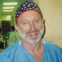
Thank you. Thanks Tamer for inviting me to come here. He’s my brother we look somewhat alike. We identified that in Brazil, so we are looking for our father, but it is okay. He is the big brother, I am the small one.
I was asked to speak a little bit about the diagnostic value of vaginal and rectal sonographic mapping, and comparing virtual colonoscopy and MRI. We are going to talk a little bit about different modalities.
First we are going to talk a little bit about virtual colonoscopy. Virtual colonoscopy was first reported in 1994 by Vining and Gelfand to detect polyps and early colorectal cancers. This is a three dimensional application of a CT-scan and these are two examples of how it looks after reconstruction of the image. You can see that you can fly into the bowel with the arrangement of computerized images of the CT-scan. You can identify the lesions also in the exam. Then in 2007 one of the first studies done was by Van der Wat. It was the modified virtual colonoscopy, a non-invasive technique, for the diagnosis of rectovaginal septum and deep infiltrating pelvic endometriosis. This study described the technique, the use of findings, but no other was presented to compare with other types of modalities like rectal sonography or transvaginal sonography or MRI. What they did is they used a 12French Foley catheter and inserted it into the rectum and insufflated it with CO2. After dilation of the bowel the CO2 ________ from the study was just eight seconds of running out the CT scan, and then they did the computerized arrangement of the study to identify the lesions. The conclusion? The findings were good for identifying lesions but more research is needed in the future. So we can compare with other modalities available at the present time.
Another study was done by Koutoukus and his group using vertical tomography. In this study they were able to identify the lesions but again, we were unable to find published data regarding accuracy, sensitivity and specificity to compare with other available modalities, but I am pretty sure that sometime in the future there will be some available studies.
Let us go now to compare other modalities, transvaginal sonography versus rectal sonography. In the review of the literature we did to bring this, since I had such a short time, we identified this study and more or less all the results are the same, so we are going to show you the result updates. In looking for deep infiltrating endometriosis, the sensitivity in transvaginal sonography was 87 against 74 for rectal endoscopic sonography. Positive predictive value was more or less the same, for accuracy it was 86 against 74. For the uterosacral ligament endometriosis transvaginal was 80 against 46, 96 against 89, 30 against 9.3, and 75 versus 50 percent in the different studies. For intestinal endometriosis it was the same, that sensitivity, positive predictive value and negative predictive value and specificity was better for the transvaginal sonography than for the rectal endoscopic sonography. The conclusion is that apparently transvaginal sonography is more accurate. It is more accurate than the rectal endoscopic sonography for predicting deep infiltrating endometriosis in specific locations, and should thus be the first line imaging technique in this setting.
Then we go to another comparison, MRI versus transvaginal sonography and this was a very good study done by Mauricio Abrao in 2007 where he compared the clinical examination versus MRI versus transvaginal sonography. You can see here he compared rectosigmoid lesions, transvaginal ultrasonography versus MRI, 98 against 83, specificity 100 against 98, positive predictive value 100 against 97, negative predictive value 98 against 84, accuracy 99 against 90, and the same with findings for the rectal cervical lesions with MRI and transvaginal sonography. The conclusion in this study? That transvaginal ultrasound is a much better tool to diagnose deep infiltrating endometriosis than was the MRI even though it is a more expensive tool.
Basing our study on the review of the literature and our own experience with 132 patients in the last year with suspected deep infiltrating endometriosis, transvaginal ultrasound is the best modality for the diagnosis of deep infiltrating endometriosis. It is an essential tool prior to surgery.
So how did we do it? We are going to show you how we did it in our practice. Dr Velez and Dr Besara here are the ones that do the surgery with me, also with Dr Fernando Rivera but he could not make it here. What we do is we prepare the bowel with a bowel prep. We put the patient there the day before, soft dieting in the am and liquid diet in the afternoon. The day of the study we instruct the patient to have three fleet enemas before the evaluation at the office. This is an essential bowel prep that will help to identify the lesions in the bowels. Let us see some here.
We always start with the upper abdomen because as you know we can find some endometriosis also in the diaphragm and other areas of the abdomen. We have a patient where we identified five lesions in the ileum and in the iliosacral bowels so we had to do the surgery to remove those lesions. We really knew exactly where we were going to look before we went to the laparoscopy. We ablated the bladder of the patient, there was no lesion. Then we did the evolution of the rectum sigmoid area here, there was a lesion. This was the probe, cervix there. This was measuring the lesion longitudinal and transfer, and transfer is very important because if it is occupying more than 40 to 50 percent of the diameter of the bowel we do a segmental resection of the lesion.
Here we have a very important slide for us. We measure from the anus to the area of the lesion so we know exactly where we are when we go to do the laparoscopy. This is one of the surgeries that we have done in this patient. We use Hennessey’s laparoscopy when we have to do a segmental resection. It was very difficult to convince the surgeons to go with us to do the surgery because in Puerto Rico you cannot touch the bowel if you do not have a surgeon in the OR. So it was very important for us to have a surgeon there. We are dissecting here, entering into the rectovaginal space, freeing a lesion here, and identifying healthy bowel so we can make the resection. We use a small incision in the umbilicus so we can put in the disc, and that is how we enter the left hand, and also when we are going to do the other part of the resection we get a bowel through that. You are going to see it right now here. This was a very big lesion, especially when we have rectal bleeding during her menstruation and a lot of pain. Preparing for the reanastomosis. This was a four-hour surgery so we do a compenion for two minutes because we know we do not have much time. This is the part where we come from the rectum. This is how it looks after we finished the reanastomosis.
In summary, we concluded that transvaginal ultrasonography is an essential tool in the diagnosis of deep infiltrating endometriosis. It should be used in all patients prior to surgery. It could prevent unnecessary surgical intervention. This is a benign, non-nicoplastic, condition often requiring very difficult surgery. A team approach is essential in the management of deep infiltrating endometriosis. Transvaginal ultrasonography should be considered when developing the new staging of endometriosis. I have been talking with Mauricio and I think we are going to do some work on that. The staging consists of two phases. First is the pre-op with transvaginal sonography. If you find the patient has a huge lesion in the rectal-cervical area or a huge lesion in the bowel, refer the patient to a surgeon that really wants to do, and is capable to do, the surgery. This is for the benefit of the patient. I had a patient with nine surgeries before we did one surgery, and we removed all the bowel lesions.
If we prepare a patient and do the sonogram and we identify that the patient does have lesions, not everybody can do the surgery. That patient will benefit from only one surgery. If you do not find anything in the sonogram then you can do the laparoscopy and do the other staging of the patient.
Thank you very much. Thank you Tamer.


