The operating room. A place not truly seen by those outside of the medical field. As a patient undergoing surgery, you’ll likely receive multiple, detailed explanations about the procedure you will be receiving. However, even on surgery day, you may get a glimpse of where your operation is about to take place, but you will likely not remember much of the operating room. As a medical student, I’ve worked with some of the minimally invasive gynecologic surgeons at the endometriosis center in Pittsburgh. I’m also an endometriosis patient, allowing me a familiarity with the room that, at the time of my own surgery, I didn’t have access to. So, what really goes on in the most exclusive location of the hospital – or the OR, as medical personnel like to call it?
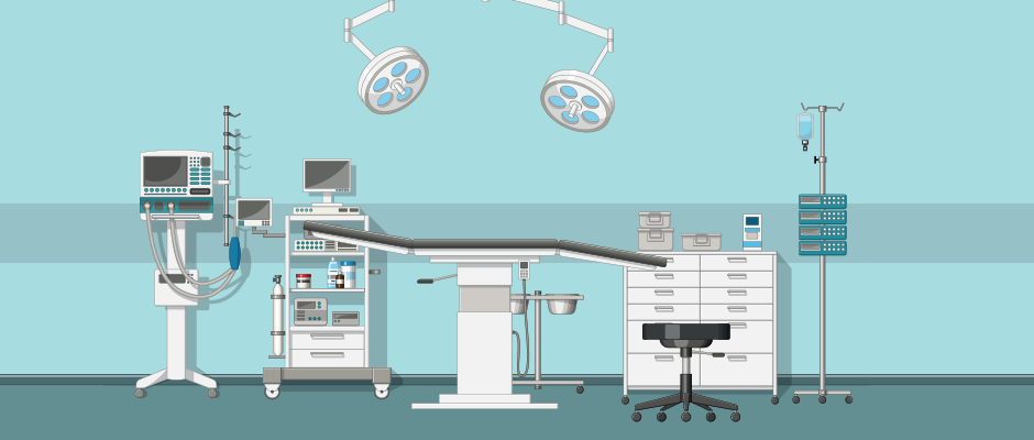
After changing into a gown, getting your IVs started, and talking to nurses, anesthesiologists, and your surgeon in the pre-op room, you’re whisked away and wheeled to the OR. Contrary to what is portrayed on television, the operating room is a bright, well-lit space, and there’s a sea of blue draping over tables, tools, and anything that is “sterile.” If you aren’t considered sterile, then you better stay away from everything blue and don’t you dare touch it. There’s a lot of equipment in the room too, but most notably there’s a lot of people in the OR. There will often be an attending surgeon, residents, anesthesiologists, scrub nurses, techs, and possibly medical students all stationed around the operating table to ensure that the procedure runs as smoothly as possible.
Once you are in the OR, the anesthesiologist will put you to sleep, and you’ll be moved to the operating table and draped for the procedure, most often laparoscopy for excision of endometriosis. Meanwhile, the surgeon or surgeons will be “scrubbing in.” This is the thorough process of washing and decontaminating the hands and forearms that surgeons need to do before every procedure in order to be sterile. They are then gowned and gloved and at this point they’re ready to operate. Just before the first cut is made there will be what’s known as a “time out,” during which the circulating nurse will likely state you, the patient’s name, your reason for needing surgery, and what procedure is about to be performed to fix it. This short pause ensures that everyone in the OR is in agreement with the plan and on the same page for the surgery. Now it’s go time.
A golden-brown antiseptic known as betadine will be used to disinfect the skin before the incisions are made. Alternatives are available if you are allergic to iodine. During laparoscopy for excision of endometriosis, four incisions are typically made: one in the umbilicus (belly button), one on each side of the abdomen in between the umbilicus and pelvis, and one a few inches below the umbilicus. Together these four incisions create a diamond shape. These incisions are also known as ports and are the sites where the surgical instruments will be inserted. One of the first things to be inserted is a tube that pumps carbon dioxide gas into the abdomen. This allows the abdomen to inflate and expand so that there is better visibility and accessibility for the surgeon. A laparoscope camera will be used for visualization of the uterus, fallopian tubes, ovaries, abdominal wall, intestines, rectum, and more. A manipulator that is inserted into the vagina may also be used in order to move the uterus around and look for endometriosis around and behind it.
How is endometriosis identified and excised? This process is difficult and requires a bit of detective work by the surgeons. They may have an idea of where the endometriosis will be based on the patient’s symptoms, but they won’t truly know until they can look in and around the abdomen and pelvis. This raises the question, what are they looking for? Unfortunately, there is not one, straightforward answer. Endometriosis lesions and adhesions may have a dark pigmentation, appearing red-black, but they can also be clear or white. There can be scar tissue in places where endometriosis has grown, and deep infiltrating endometriosis may appear nodular. Some lesions are not seen because they are behind the peritoneum or within ovaries or other organs. When these are more than 5 to 10 mm, they can be found on sonogram or MRI.
Because you cannot rely on appearance alone, it’s important to have a surgeon who has extensive experience and training in identifying and excising endometriosis. As a medical student who has had the opportunity to observe an endometriosis surgery, when I asked how endometriosis is identified the excision specialist responded, “sometimes you can feel it.” Once again, this reiterates how complex endometriosis excision is and how surgeons who are well-versed and knowledgeable about endometriosis will be able to identify endometriosis lesions not only by looking and using a surgical camera, but also by feeling the differences between different areas of tissue in and around the abdomen and pelvis. Thorough removal of endometriosis is imperative for positive patient outcomes. As Dr. Seckin puts it,
“An endometriosis lesion is like an iceberg with just the peak being visible while the actual bulk of the lesion is still within the underlying tissue. The underlying part of the lesion can regrow again leading to recurrence of symptoms. Therefore, complete excision of all visible endometriosis lesions is essential for full symptom alleviation and to prevent a recurrence.”
It's especially important to note that excision surgery fully removes a deep endometriosis lesion while electrosurgical or laser ablation surgery, where a surgeon burns the lesion off thus leaving behind its root (the majority of said iceberg), does not. Although laparoscopy for excision of endometriosis is most often an out-patient procedure, the complexity of identifying, removing, and diagnosing this chronic illness involves an intricate operation. Experienced excision specialists and continued advancements in treating endometriosis are imperative to ensure success for each patient in the operating room.
During laparoscopy for endometriosis, it’s common for multiple types of procedures to be performed in addition to excision, such as ovarian cystectomy or ureterolysis. An ovarian cystectomy is the surgical removal of cysts on one or both ovaries. This is necessary when there is an endometrioma, or a chocolate cyst on the ovary. This type of cyst is filled with endometriosis and endometrial-like tissue. During surgery, the wall of the cyst will be cut and the contents of the cyst along with the cyst-sac itself will be suctioned out through one of the small incisions that was made earlier in the abdomen. Ureterolysis may also need to be performed if endometriosis lesions are found on the ureters which are the tubes that connect the kidneys to the urinary bladder. This procedure aims to expose and free the ureter from the adhesions.
Endometriosis is often a multi-organ disease, growing on the uterus, fallopian tubes, ovaries, bladder, ureters, and rectum. To further complicate things, endometriosis has been found to grow on organs outside of the abdomen and pelvis, including the intestines, diaphragm, lungs, and brain. Therefore, there may need to be additional surgeons from different specialties on standby in addition to the endometriosis excision specialist. Urologists, gastroenterologists, and thoracic surgeons could provide added expertise if needed, depending on where the lesions are located.
Once the endometriosis is removed, the incisions will be closed, and any tissue excised will be sent to pathology in order to confirm the presence of endometriosis. The patient will be taken to the PACU, or Post Anesthesia Care Unit, until they wake up and are cleared for discharge.
So, when you wake up in recovery, everything may feel like a blur. You have no sense of how long you’ve been asleep and are eager to know how the procedure went. However, it’s not often that patients actually see, let alone talk to, their surgeon after surgery in post-op. In most cases it’s up to your nurses or family members to pass along the news of how the operation went. After being monitored for a few hours in recovery, you’ll be discharged and sent home. And just like that all the fear, worry, and nerves you had leading up to surgery are gone.
Now starts the healing process and hopefully the start of a better life with less pain.