International Medical Conference
Endometriosis 2024:
Elevating Sampson’s Century Legacy via
Deep Dive with AI
For the benefit of Endometriosis Foundation of America (EndoFound)
May 2-3, 2024 - JAY CENTER (Paris Room) - NYC
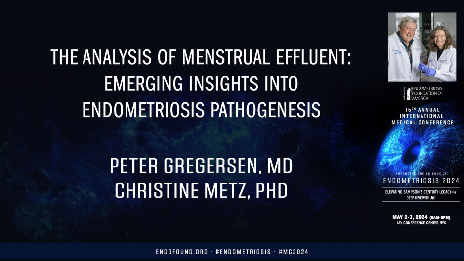
Okay, so we started almost a decade ago picking up from work that many others had done on stromal cells that are from biopsies and showing that they had a defect in residualization. We wonder if we could demonstrate that in menstrual blood's, derived endometrial stromal cells. And in fact, we were able to do that in spades. And we got very excited about this because we thought, oh, this could be a diagnostic or at least a screening test for endometriosis. And while it could be, it just doesn't lend itself practically to doing that, it's very complicated to get cells, treat them and measure all these responses. So while it really looks good on the RO seeker in terms of practice, it's not so great. I will also point out that the published data from biopsies is not quite as striking as this. And I think the reason for that is because we are looking at cells that have not only gone through the luteal phase, but they've also gone through menstruation. And we're beginning to think that these abnormalities that you're going to hear about really are related to what's going on during the luteal phase, but also the effect of menstruation.
I, yeah. So we spent a lot of time figuring out how to collect menstrual effluence. Could it be shipped? Did it need to be on ice? How did it have to be dealt with? And one of the amazing things about endometrial stromal cells is that they are survivors. So you don't actually have to worry about the stromal cells per se very much because they survive. And that's why a lot of our work has been on stromal cells. Okay, somewhere in the middle of this, we were growing stromal cells and sort of looking at the insulin effluent, but didn't really grasp what probably all of you knew, which was that the menstrual effluent is full of tissue fragments, not just free cells. And so this is just an example of an h and e of menstrual effluent showing that you actually see glands, you see s stroma that are intact, they're organized somewhat differently or maybe not as well as they are from a biopsy, but nevertheless, they're there to be studied.
So we decided that we wanted to know about all the cells in these fragments. So we undertook single cell RNA sequencing of this. I won't go through a single cell, and this is just the data is published. But that just revealed to us that endometriosis menstrual fragments are totally different than menstrual fragments from controls. And they have a lot of abnormalities that we haven't still fully understood, but they clearly have a depletion of uterine NK cells. This little group up here are proliferating uterine NK cells. They're nearly absent in the setting of endometriosis. There is an increase in various lymphoid cells, particularly B cells, but actually T cells show enrichment as well. Then in terms of numbers, the numbers of stromal cells are not different, which might seem odd because one of the defects is deci visualization. But smal cells are dely defective digitalization because of what they're like.
So just again, this is a waterfall plot of the numbers. Lots of unit NK cells in controls, lots of BET cells in cases. But if you look at the subset of stromal cells, you find out that there are a bunch of subsets within the stromal cell compartment. One of them is a IG FPP one positive cell, which of course is associated with de digitalization. And those subsets are in fact enriched in controls. Whereas several other subsets that we call IL 11 and MGP positive G-protein positive are enriched in cases very, very significant P values here are 10 to the minus 15. So they're very, very different. And if you look at what these different subsets make, they make things that are associated with inflammation. Not surprisingly, they're estrogen responsive disease. And thing that's gotten us intrigued recently is that these cells express markers that suggest cellular senescence, which is a very difficult thing to prove. And we'll talk about that a little bit. So, whoops.
Oh, we just did that. Isn't there supposed to be something else in there? Anyway, this led us to this concept that the stromal cell differentiation is skewed normally towards good ization, but in the setting of stress, inflammation, maybe the inflammation of menstruation contributes to what we find. Chronic endometritis, which I'm sure you're aware is associated with endometriosis, progesterone resistance, which is quite well described. This digitalization event is clearly dependent on progesterone. So we felt that probably this change and what these cells end up doing is what leads to these changes in single cell RNA-Seq, and that we believe that pre senescent cells are part of that. Pre senescent cells are very interesting because they make a lot of pro-inflammatory markers. The uterine AK cells, as I've told you, are low. And in fact, there are others that have shown that they in fact kill senescent cells. So we wanted to replicate this data, and I'll just show you quickly that in fact, this data replicates in a prospective project where we get menstrual effluent several months prior to surgery, and then at surgery our endometriosis is confirmed or not. And then we compare, in this case, we compare controls.
Why is this doing it? We compare controls to these new prospectively collected cases, and we see exactly the same waterfall plot, low uterine NK cells, low uterine NK cells, they're proliferating and high B cells, T cells, and no difference in the number of stromal cells. But the stromal cells, in fact, are different as we saw before. So if we remember, we had an enrichment of stromal cells that produce IG FPP one in the original data, and we see that again in this prospective data. So we're pretty confident that these phenotypes that we're seeing in menstrual blood are reproducible and go with endometriosis even before the diagnosis is made. Many of the people we saw before had had a previous diagnosis. So again, three things decreased residualization Jesus, why is this doing that?
An increase in inflammatory immune cells and altered stromal cells that suggest a senescent phenotype and decrease uterine NK cells. So more recently we've tried to take a new approach at identifying these phenotypes using spatial proteomic imaging. We're using lico cell type. There's other methods out there. Koya is one of them where pathologists are now using this to not just stain one or two antibodies in the tissue, but stain 20 or even 50 antibodies all at once. You can do this in an automated fashion with this machine, which is quite expensive. So there aren't that many around.
And we've been working on this for I would say eight months. And this is not a trivial, it's not for the faint part. First of all, not all the antibodies you want are available from Leica. The protocols for preparation of these FFPE samples, you really have to work at. There's a baking step, there's a dation step. You got to have 'em stick on the slide so they don't wash away when it goes through the thing. A lot of mistakes were made getting this to work. It's now working. There's also a pretty complicated set of imaging software with a steep learning curve. And in fact, we have two of the people that work on this are in this room. One is a biologist and one is a informatics expert. And it really takes those two people looking at these images and doing all the controls for background before you can really be confident of what you're seeing. And the data storage is enormous from these images. A terabyte in experiment is low, so you have to have a lot of backup. So I just want to show you a few examples of this because we don't have a lesson on the basis of this yet, but this is a lesion and a very active lesion stained with about eight antibodies. You look at this, Hey, why is it doing that?
What is this the play? Yeah, I'm going to do the play. Click here, I'm doing it. Okay, so you look at this,
This is I think about eight to 10 antibodies. There's a lot separate them out. Cells. Cells,
The stromal cells. You can separate them out and ask the machine questions about how many there are, what's next to what. So this is just poised for ai. And I'll just show you that You can see the same thing using stro endometrial fragments in me using the same approaches. You can see a lot of these markers altogether and separate them out. And we're just at the beginning of stage of this. We've gone through what, eight or 10 rounds of this with maybe 30 antibodies. We're still collecting antibodies. You have to validate antibodies. So hopefully there will be an accumulating set of antibodies that'll be useful for this that will be able to release to people. So Christine is now going to tell you a little bit more about how we think we're making sense of this in terms of disease fat attacks.
Okay, so I'm going to pivot a little bit and share some of our data that embraces Samson's theory and supports targeting senescence in fibrosis for possibly treating endometriosis. As everyone in this room knows, there is a lack of effective tolerable medical treatments, pharmacologic treatments that is for patients with endometriosis. Most of these agents treat symptoms such as pain and do not halt. The progression of disease fibrosis is a really intricate part of endometriosis. It's often a major component of the lesions, pushing out the stromal cells and epithelial glands, making diagnosis even more difficult. Fibrosis is not included in the A SRM or NZN classifications and fibrosis is not targeted by any of the endometriosis therapies that we have today. However, many lesions contain active fibrosis. And in fact, as the disease progresses, you often have increasing fibrosis. And fibrosis is just a characteristic feature that we now propose that senescence underlies the fibrosis in endometriosis in some patients, and that could be leveraged for diagnosing endometriosis and treating it. So as Peter said, we've done single cell RNA-Seq on menstrual effluent tissues, and we see major differences.
We see major differences between cases and controls with a decrease in the uterine NK cells and an increase in the CD four T cells and B cells. And we also see that this RNA-Seq data supports a disease model for us to interrogate endometriosis. And I'm going to focus on the bottom half of this slide where there is an increase in senescent cells, which by definition are growth arrested and they resist apoptosis, but they're not simply innocent bystanders. In the disease model, senescent cells actually secrete what is known as senescence associated phenotype, secretory phenotype markers such as extracellular matrix chemokines, cytokines, and other inflammatory mediators that drive fibrosis creating a vicious cycle. There is very good evidence of senescent cells in the endometrium, and people have shown that uterine NK cells, which are depleted in patients with clear these senescent cells in order to promote fertility.
We would also suggest that or speculate that there are additional alterations to menstrual cells delivered to the pelvic cavity by their presence in menstrual blood. These cells are bathed in red blood cells and exposed to iron. So based on some work in the setting of renal fibrosis, a paper published by moss in 2023 reported that iron accumulation drives fibrosis and this SAS phenotype or senescence associated phenotype. And these senescent cells actually persistently accumulate iron even after an iron surge. And these high levels of labile iron create an increase in reactive oxygen species and the SaaS phenotype. We suggest that this iron is a fuel to these senescent cells which are delivered to the peritoneal cavity. So as Peter said, the endometrial stromal cells in patients with endometriosis have enhanced IL 11 and end GP clusters. Those two clusters are very associated with pro fibrotic and pro senescent phenotypes.
The major differences that we see in menstrual effluent in cases and controls can all be linked in one way or another to fibrosis. This increase in B cells has been shown in fibrotic lung disease and antifibrotic treatment actually alters B-cell function in that condition. The decrease in the uterine NK cells could be linked to the depletion of NK cells associated with an increase in liver fibrosis and actually antifibrotic activity of NK cells in that condition. And then of course, with the change in the CD four T cells, those cells are known to mediate fibrosis in non-alcoholic fatty liver disease. And senescence has been shown to mediate fibrosis in pulmonary and renal diseases and lytic agents, which Dr. Nimeroff referred to actually improve patient outcomes. So with all of the flurry of activity in anti-aging, there has been a big development in seno litic agents that take out these senescent cells.
So just to remind, senescent cells resist apoptosis, seno litic drugs targets scs scaps are the senescence associated anti apoptotic pathways that allow these cells to resist apoptosis, and that's what the seno litic drugs target to mediate apoptosis or cell death. And two s alytics have been used in many clinical trials as well as preclinical trials, quercetin and dasatinib and both renal fibrosis and pulmonary fibrosis have shown positive outcomes in patients. We focused mainly on cetin. It is a natural flavonoid and considered a generally recognized safe agent. It is anti-inflammatory, antioxidant, anti almost everything as well as a lytic agent. And we've started to do a lot of experiments to look at its effect on endometrial stromal cells and to verify that in fact it had an antiproliferative effect, which Dr. Nimeroff referred to. And indeed it does. And in addition to its antiproliferative effect, quercetin enhances de digitalization of endometrial stromal cells in patients who were controls as well as patients who had endometriosis.
Quercetin also reduced the production of SAS mediators, specifically in this case IL six and MMP three, which are known to block de digitalization. And we've done many more experiments, which we have not enough time to talk about today. But I could go through what we think is happening here that quercetin alters the irk one, two and a KT signaling pathways, which are both associated with increased proliferation within the endometrial stromal cells. And when those two cells, those pathways converge on P 53, which would inhibit apoptosis in those cells. And in the presence of quercetin, it is able to block those two signaling pathways and actually promote P 53 expression, promote P 53 phosphorylation, and increase the apoptosis of what we think are senescent like cells in the menstrual effluent. So overall, we believe that quercetin may be used as a treatment. There are several papers that support its use in animal models.
There are several papers that support its use in pain, including cancer pain and patients neuropathic pain and other forms of pain. So we're very excited about this possibility. And for us, what's next is a quercetin supplementation trial. And the goal here would be to determine the effect of quercetin supplementation on stromal cell function, ex vivo, we would provide quercetin supplementation after patients have already given us, I should say controls, healthy controls have given us their menstrual effluent samples. We would actually begin the quercetin supplementation around the time of ovulation, and the treatment would go through day one of menses. And on day two of menses, we would have them collect their menstrual effluent again and with the pre and post menstrual effluent collections, we would look at decentralization in those cells as well as do single cell RN aeq to see if we see any of the alterations that we might expect with quercetin treatment. So I would end here and say many, many thanks to all of our rose participants. Special thanks to the endometriosis foundation who actually gave us the first grant that we ever had to study, menstrual effluent, our clinical collaborators, our nonclinical collaborators, and everybody in the lab who has made this team work. Thank you very much.


