International Medical Conference
Endometriosis 2024:
Elevating Sampson’s Century Legacy via
Deep Dive with AI
For the benefit of Endometriosis Foundation of America (EndoFound)
May 2-3, 2024 - JAY CENTER (Paris Room) - NYC
Good. Okay. I got more recognition from operating without a mask than I ever did with a mask. And really, for most of my career, we did not use a mask. I mean, we started doing laparoscopy with our head in the surgical field. So what use was a mask? So anyway, that's the way it was, and I like to say hi to Fili and Anastasia, but Anastasia couldn't make it. So anyway, I'm from the coal regions of Pennsylvania. I'm from the area where most of the mafia that you hear about, like the Irishman, all those stories come from there. And my mother being a physician delivered most of them. So that gave me my background. My background in endometriosis was from Robert Kissner, who was the major person in Boston and in much of the United States. He wrote extensively. And as you see at the end of that slide says, what was a cul-de-sac?
They had no idea what a cul-de-sac was. They did beautiful surgery on the ovaries, beautiful surgery on suspending the uterus out of the pelvis. But the cul-de-sac still stayed, remained with endometriosis, and kisser was a good surgeon. He could do gallbladders, he could do the whole gamut of general surgery. He could do bowel resections, but the cul-de-sac, they forgot about laparoscopic expertise. Where I'm from was very hard to obtain. In fact, when I started practice, I had much laparoscopy. I was trained as a vaginal surgeon, and my main interest was vaginal surgery, not abdominal surgery. And within four years, I had done it, 500 laparoscopies, mainly for sterilization, because the area I went to, no one did sterilization, laparoscopies, and most of 'em did not ever use laparoscope for fertility evaluation either. So anyway, in 1976, there was no one to learn from. So the only tool we really had was the kleger bipolar forcep. So we could control bleeding.
Early on, I discovered I could do an ectomy B laparoscopy, and that was back in 1976. And when I tried to publish it in 1985, they said, it's too radical. Forget that. So now we're back in bipolar and doing that stuff. So anyway, laparoscopic. I like to show this slide here. This is the way we used to operate. We did not have video. And even in this picture here, you could see the thing coming from the laparoscope. It's a beam splitter. So even when video came available, I didn't believe in it. So I thought I could do much better with my eye than a video camera. So most of the cases, even into laparoscopic hysterectomy, they were done with eye, using my eye, not using a video camera. So anyway, going through the different procedures that we did, I like to emphasize that 1983, I made a decision not to do any more laparotomies.
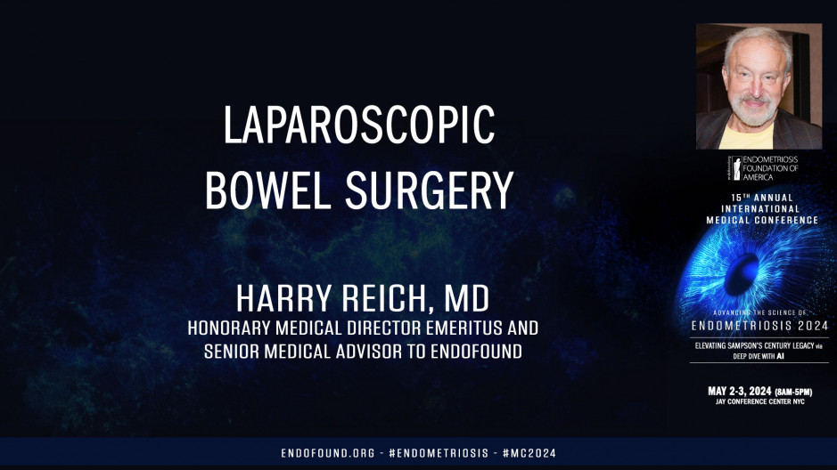
I pretty well stayed to that for the rest of my career. Most procedures we could do by laparoscopy and vaginal surgery. I like to say I am indebted. I did spend two weeks, or I'm sorry, two days with Bruja. And the main guy I really learned from was Uber Monas, Uber Monas, the first ectopic pregnancy in 1974 in, he called it a caesarean section on fallopian tube. Anyway, so he was the man who got behind the whole movement, I think in France. Kurt Sim, I didn't really pick up much from Kurt Sim in my career. The only thing I picked up from him was a concept of suturing. And he says, rich, if you learn to suture, you be king. So I did learn a suture. Anyway, 1985, I started at A GL. I presented artwork with ovarian endometriomas and with sapa ectomy. And we started right after that presenting about the cul-de-sac and doing the cul-de-sac dissection. And in fact, I had a debate for the Endometriosis Society with Robert Franklin, who's a very famous guy down in Houston, Texas. I'm doing this stuff. Anyway, I want to get to bowel surgery, which is a topic of my presentation today, and hopefully my videos will work. Now, the first one I show, I want to go, let's do this to halfway through.
See?
Can we go to halfway through?
Sure.
Okay.
Like this? Yep. That's going to take time.
Nope, you wipe it out.
Fuck up. Hopefully it's going to work, huh? Hopefully
It's not going to work.
Yeah, it's like
Really videos, huh? I'll show the whole video if we could stop it. Okay. Okay. This was the extensive endometriosis operation, and the reason I show you this is to show you what endometriosis nodule of the rectum really consists of. So when we get that, so first of all, one has to dissect the uterus and the vagina off the rectum, and I would go straight for the vagina or straight for the cul-de-sac in the middle. And most of the Europeans I know go down the sides and dissect the ureters first. But what I want to show you there is there's a nodule in this rectum, and right now you just see a rectum.
But we start with a CO2 laser, and you could see the precision of the CO2 laser because I'm working on the deep fibrotic endometriosis junction with normal appearing tissue. And what I do is I go round and round in ever increasing concentric circles around the lesion until I dissect out a big marble, a big nodule, and I'm really working at the junction of rectal mucosa without entering the mucosa, the mucosa layer. And I'm dissecting the whole nodule from surrounding muscle, and it comes out as a big nodule in the rectum. Again, it'd be much easier to just resect the rectum, but that's not what we're doing at this stage because I wanted to show what a nodule really was. And to do this, identify the deep fibrotic endometriosis, which is white. It's not a mystery. It's white. It is not DIE. It's deep fibrotic endometriosis. There's nothing invasive about it. It's caused by many years of inflammatory reaction around the endometriosis implants. So I finally get the whole nodule dissected out. I can then take it off the mucosa of the rectum, and you'll see when I put the rectal probe in, it's right there. I'm right on the rectum. On the mucosa.
The CO2 laser was marvelous to be able to do this type of work. Again, I use micro bipolar force where I irrigate and see exactly where the small bleeders are. And just by touching the bleeders, I'll stop the bleeding and get complete hemostasis.
I
Thank you, tar. That worked out well. Do you
Want me to move?
No. Here, I'll just move it
Forward.
Yep. Next slide. Yeah,
Sure.
I just press
This. This
One? No, right to the next slide here. I just, that's it. That's it. Okay. Now here you can see you want to play this? Play it. Yeah. Here, I just want to show you that you can use electrosurgery with only cutting current electrosurgery, which acts just as C of two laser. The play.
Interesting. It plays very slow, but it's happened. Your
Computer, well forget it. It's just a slide to show that you could do the same thing with electrosurgery. Here, I'll just go to the next slide. Okay. My time is going, so I need to go to the next slide. Bad.
I think it's stuck all
Day. Stuck again. Yeah.
Stuck in there.
Some of these are old slides. Okay. Get past there. This one, right? Okay. Now I'm back. Okay. Beside, okay, I want to go stuck again. Stuck
Again.
Go back next forward. I want to keep going to the next slide. Perfect. Yeah. Best.
Interesting. It's got stopped.
Okay. I want to tell you a little story. In 1989, I went over to Wickham, who's a urologist in London, and I met this Dr. Gerhard Woo from Germany, and he was doing rectal resections through an anal scope. So I said, my gosh, if he could do that, it should be very easy to be able to resect rectum by going inside the rectum instead of looking on the outside. So we started doing this procedure right after that in 91, where we would, with an rectal nodule, insert a circular stapler, as you could see inside the rectum, push nodule into the circular stapler and excising. And we did about 60 of these, A very nice procedure here. You could see with the endometriosis split open instead of a nodule like you saw in the first video, we can then exert the stapler, excise the lesion, and then carefully you could see taking the lesion out and underwater exam afterwards, which we always did for all our procedures, and worked for a lot of time until we got complete hemostasis in the cul-de-sac. You could see the lesions in this case, and there's the type of tissue that we would get. Now, what do you do when you have this type of lesion? This is a sigmoid colon way up there, sigmoid. So I said, well, I'll resect it just like I did the cases I just showed you. So I start and I said, start this video.
We'll play it right through just the way it is. So I start and I find she has a rectal stenosis, and you can see I'm stuck. I can't move my rectal probe forward. I'm stuck at the stenosis. I have to get all the way up to the sigmoid. Now I have to tell you, beginning of the operation, I put a suture, I dissected out the sigmoid, and I put a suture around the sigmoid to avoid rectal spillage coming down into that area. But anyway, so here, because there's a stenosis, I excise the endometriosis on the anterior rectum. One can see the stapler in the rectum. I'm not going to be able to use it at this juncture, that's for sure. So at this point, I'm just debulking endometriosis, deep fibrotic endometriosis, not deep infiltrating endometriosis. And you could feel it. It doesn't feel like normal tissue. You could feel the crunch and your instrument makes a crunch sound as it goes around the lesion. So once I have the proximal stump pretty clean, I start mobilizing the sigmoid. Now realize I operate with two lower quadrant incisions, two fives, only fives. I have a scope in the umbilicus, and I have two fives, one on the right and one on the left. No, third, I've done that all my life. I sort of had very strict rules from the early days, and I didn't change them. And we mobilized the sigmoid.
Every now and again, one must use a bipolar forcep Early. It was just monopolar cutting current. I do not use coagulation current ever through my monopolar devices. I just use cutting current with a blunt tip. You could coagulate with the bluntness of it, or you could cut with the sharp edge of it. So we then proceed upward until a sigmoid is mobilized. Remember, this is the main lesion. Here is a sigmoid lesion. You can see the mobilized sigmoid being pulled out. Oh, again, just bipolar. I'm sort of limited because they only have the scissors that does not have any electrical attachments to it. And we make rapid instrument exchanges when we need to get to the bipolar. My surgical assistant, my nurse would hold the uterus up with her belly button, so she had two free hands to be able to hand the serum. Now here's the mobile is sigmoid up to the lesion, and then we grasp the bowel. At the cut end, we pull it down through the anus, and here you could see the nodule. You can see at the end of it, the nodule that we were after of the sigmoid.
So then I divide the sigmoid at that juncture and put the anvil in, and then connect the animal after a purse string suture around the distal end of the colon. We bring together at the end, like I showed you earlier, we always do an underwater examination to check for hemostasis. And I fill the rectum up with blue dye, with indigo carmine type thigh, and I could usually see if there's a thin out area with a blue dye. And that's it. Let's see. Next slide. Yeah,
Next slide. Just a second.
Okay, just about done
If I
Can get it. There's only one more slide. Two more slides. I have two more
Slides. Yeah, I don't know what to
Do's. Another slide after this.
Okay. Yes, this is the one.
Okay. Okay. Okay. So we check underwater. Underwater exam,
Simple adhesion reduction, ringers. Lactate, 2000 ccs. That's what we used from the early eighties onward throughout my career, and resulted in very few infections. And I'd just like to close with the emphasis on the beginning of these operations for any gynecologist practicing Today, you'll see all this literature that we shouldn't do rectal vaginal exams. That's crazy. I mean, a rectal vaginal exam is the most important part of your gynecological examination because you could 10 up the cervix, you could feel the rectum, and you could feel the uterosacral ligaments. It could make your diagnosis very nicely. Thank you very much for your attention.