Endometriosis Foundation of America
Medical Conference – 2012
The Technological Transformation of Surgery: Film & Medicine
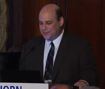
Steven Palter, MD
He will talk lots about films and I think somebody is going to come out and award somebody for cinematography in medicine. In any case, let me tell you a little bit about Dr. Palter. He is the founder, medical and scientific Director of Gold Coast IVF in Syosset, New York, and the former Clinic Chief of Reproductive Endocrinology and Infertility at Yale University School of Medicine. He founded and is the Program Director of the Center for Excellence Program in Gynecologic Minimally Invasive Surgery. Dr. Palter’s passion for technological innovation is well known and he performed the world’s first HD and 4,000 surgeries. It is a great honor to introduce Dr. Palter.
Thank you so much for that kind introduction and thank you to my friend Tamer for the invitation to be here and my colleagues and friends who have spoken to you before.
As mentioned, I am a reproductive endocrinologist. I am a fertility specialist but I work in multi-modal therapy; surgery, IVF, whatever we need to accomplish our results for patients, either to get pregnant or to maintain their fertility. What I am going to talk to you about today is to take the introduction that Dr. Setubal gave you of where how film and medicine set the stage for this collaboration and then show you what came next. How we moved through the 1900s to where we are today and to give you a vision of where we are going to go in the future.
I am not going to talk to you so much as a data driven lecture but I want to give you a vision of how the marriage between technology and film has changed surgical practice and I want to show you why everything that you do today will, within the next decade, be really obsolete. And why instead of that being an area for fear, you should see this as perhaps the greatest opportunity for change and transformation of care of our patients and how you perform your surgical practice that we have ever had. I am going to talk to you about the innovations in cinema and their impact on medicine, why laparoscopy has fundamentally changed the mindset of the surgeon, and share a vision of how technology transforms surgery.
What I am going to give you now is a visual journey starting from where Dr. Setubal was. If you look back into the 1900s to 2015 I think what we had was, call it a continuum of interactivity. You basically had a picture on a screen and you brought it in and then the audience sat there. I will show you in my first videos, also rescued from some of the US archives, is how the image got better and better approaching what you could do with your eyes and your hands. In Hollywood with some of the people that we collaborate with and in video technology we increased the realism and sound and color. But where future technology is going to go is beyond both immersive reality and beyond the natural abilities of the surgeon. Where instead of the technology just being a tool to approximate what you do it will actually enhance what you could ever do. Antonio showed you the Lumière brothers and he showed you this film or the first film that was shown in the Paris Grand Café. The point I want to show you is that the reaction of the audience was remarkable when this was shown. At this point in the movie the crowd was 35 people or so, three quarters of the audience stood up and ran out of the room in terror because they were sure that they were going to be crushed to death by this oncoming train. In their world set in their mind they had never seen an image of a train coming at them on a screen. There had never been a motion picture on the wall and so to them this seems so real that they were sure that actually a train was coming through and they would be crushed despite when you look at it now it seems absurd.
I am going to show you why, in fifty years, people will look back at what we are doing today with the same mind set, “My God, how could anyone believe that these were images? This was the most crude and rudimentary things that we had ever seen”. Let us talk about the marriage of endoscopy and film because nowhere more in medicine than endoscopy have the two been together. This is a film that we rescued from the American National Archives in the National Library from the 1930s. This was the first coupling of an endoscope to a camera. Look at the size of that monster. It was a film camera and it was right at the time when “Gone With the Wind” was coming out. In the late 1930s they basically used this just as Dr. Setubal showed you to educate and to show people how procedures were being performed. This is one of the first uses of this. This is a, by the way they are using cocaine as a local anesthesia, that is what they used at that time. The patient is blindfolded and they say you have to restrain them and here they are recording the film for teaching abilities. Again, no one had ever seen something so realistic as this. In fact there was a backlash saying perhaps this is inappropriate to show something of such real candor in a public meeting.
It then made it to this point, this was rescued from one of the German archives and again, we have switched our machine so much – hopefully they will all work. This was rescued from a German archive and this was the first laparoscopy that was recorded. You will see the patient is strapped down when he comes in. The technique is quite a bit different than we use today. They used room air with a bellows to insufflate the abdomen. What the commentary in German said in this is they are remarking at the “miraculous image that is akin to what you could see with your naked eye”. They were performing biopsies of peritoneal tumors to confirm the diagnosis of cancer.
In 1971 this was the first from Upjohn, the first laparoscopic teaching film. The same commentary: “…laparoscopy is indicated in cases where routine diagnostic procedures fail to reveal the cause of acute pelvic pain and for tubal ligation. It is especially useful in differentiating between acute pelvic inflammatory disease, physiologic ovarian cyst and tubal pregnancy”. In 1971 that was the state of the art and when that film was shown at the AAGL National Conference people suddenly realized that you could do things with video that would be akin to your eyes since the video was as good as your eye. But what I want to show you is how the marriage of technology enabled things to happen that previously would have been impossible.
This was a film that I found in the US National Archive that was labeled “Miscellaneous Surgical Video”. What it actually was, was Patrick Steptoe of Steptoe and Edwards fame, who invented in vitro fertilization performing the first procedure where they did the first egg retrieval that he said was possible because of laparoscopy and his first attempt at getting an egg. You will see the equipment that they used. “The examination of the abdomen by means of laparoscope has proved to be a most valuable diagnostic advance. One of the most important recent developments is the recovery of oocytes after priming the ovaries with gonadotropins. First of all the ovary is held steady in position. A follicle is punctured and the contents are aspirated at a low pressure so as not to damage the oocyte”. There is the first recorded egg retrieval before IVF…
Now I had the pleasure of meeting Steptoe and Edwards, and Edwards said, and I will quote, “I read about this chap Steptoe and his work with something called a laparoscope. He was writing about how he had managed to reach the fallopian tubes and I thought if he can do that he could reach the ovary and we could invent this new procedure”. It was the ability to have the technology to perform laparoscopy that gave them the idea. He actually watched Steptoe do a video of laparoscopy and he had the idea that IVF could come.
Well, where did this come from? What was the reaction of the traditional surgeon when he saw his first laparoscope? I mean Harry tells us stories of when he performed these miraculous, wonderful procedures that cured the patient, and they said, “You must be insane, we’ll have to put you in jail for doing this”. Kurt Semm had to have a CT-scan of his head because they said, “You must have a brain tumor to think of doing this procedure”, even though it actually worked. So what uses did the first laparoscopic surgeon imagine and where did the technology come from? I guarantee you that when this technology was invented no one had the beginnings of a thought that we could do anything but look inside. It actually was the same path that occurred in technology outside of medicine. Something that I am going to show you is how progress is ever accelerating. How the rate of change is accelerating in the world and the difference between 1970 and today will now occur in just a few years.
This is Boyle and Smith, they worked in Bell Labs in 1970 and what they are showing you here is where they invented the CCD, the charge couple device, which was the chip in our cameras. This camera you see that broadcast is the camera in the operating room. They won the Nobel prize for this and what they were trying to invent was a video phone. They were not trying to do anything for surgery but then the surgeon thought perhaps we could couple these technologies. This is something called analogue transfer. You take a technology from another field and you bring it into your field to change the mind set of how you do things. Now just think about how fast technology is changing. Look at your cell phone today versus where it was five years ago. The same thing is going to happen with our surgical tools, because, in fact, this was the first VCR. That is Ray Dolby with his Emmy that he won for this. It cost $50,000, it was the AMPAX 1956 year VR1000, and it cost $50,000. The only use of it that could be imagined to invent recording was to record the news and Douglas Edwards was the anchor and to show it later, a few hours later. There was no thought that anyone could come up of why you would ever do anything with this. But look how once the technology existed, new uses, new procedures and new medical uses came out of it.
Medical endoscopy is an outgrowth of commercial broadcast equipment. Film, video and medicine were married and this is Camran Nezhat with the first camera he used. It was actually hung from a boom in the ceiling because it was so heavy. I will show you a little bit how we took that idea to do this project again today.
This is the first course from the first series. This was found in an archive in Hawaii in the early 1970s where they said video would be for mentoring. This was the first resident training. This was the view of the pelvis. And what they comment on is again this is the most remarkable realistic image that they had ever seen in their life. Nothing could be better than this and they said, “Look, this is the first time we’ve actually had a color image”. The cameras used to be black and white prior to this. They used the black and white cameras to get increased resolution and then they switched to the color and again that is the camera that is being used. When we look at this now, you realize this was 1970, so it is 40 years ago, this is not 100 years ago this is 40 years ago. When you look at this in the room it is incomprehensible to you that this could have been in your lifetime when it is so utterly primitive and poor quality.
Let me show you the theory I have of the “technological transformation of surgery”. Thomas Kuhn in 1962 wrote a book, “The Structure of Scientific Revolution” where he coined the term “paradigm shift”. That is thrown around a lot today. What it means is that when something occurs that shakes up your whole view of the world, such that it not just gives you an idea but your whole system of doing things no longer exists. Changes will occur in your entire world view. What I will show you is that laparoscopy is that point for all of medicine. Since antiquity the surgeon’s mind, there was a direct interaction between the surgeon’s hand and the patient’s body. There was an incision made, the surgeon stuck his hand in just as Doyen did and he cut something out. I will tell you from 1700 to 1970 very little changed. It is the same idea. You made a cut, you put it in, and all that changed was the suture, the antibiotic and the clamp. But there was no radical change in how things were done. You were guided by your traditional senses and feedback. So the OR of 1950 was no different than the OR of 1850. The laparoscopist’s mind completely changed that because for the first time now there were remote instruments viewing the procedure on a TV monitor. In my theory laparoscopy is a critical pivotal point where the surgeon no longer is directly interacting with the patient but there is technology between them. What does that mean? It means that surgery can be fundamentally transformed because a knife cannot change very much. A clamp cannot change very much. But microchip technology follows Moore’s law and it increases exponentially. Once you put computer chips between you and the patient suddenly there are no limits to how procedures will change.
The interaction between the surgeon and the patient through a mechanical and electronic interface is a fundamental paradigm shift in medical care. It sets the stage for technology could then modify the interface. This was Colonel Christoph Kaufmann working in DARPA where they made the first robotic surgery. This was the precursor of the da Vinci. So, if technology is between you and the patient then a robot could be there. You could, in fact, simulate the interface. If technology is between you and the patient you could eliminate the patient and reproduce the surgery for training.
Has endoscopic surgery maxed out? Have we gone as far as we can or can we reinvent surgery? Now I have brought you up to the point where we are now at 2012. Let me show you where I think we are going to go in the next ten to 20 years and how I believe we will reinvent surgery.
In my technological theory of medical progress for the 21st century the digital revolution is now upon us in medicine. I showed you how technology is in the interface and we have now entered the exponential growth phase where I will show you why I believe that we will have and order of magnitude ten years progress in one year as we move ahead. We have mathematically modeled medical progress in surgical innovation and compared it to what is happening outside of medicine and showed that the same rate of rise that is occurring with your cell phone will occur with surgery now that there is technology involved. Moore’s Law said that computer power the number of transistors on the chip would double every one to three years and it would be at a lower cost. Basically technology is not 30 times less than it was 30 years ago, it is 8000 times less. Moore looked at processing power and he said, remember this is a logarithmics scale, that it is an exponential rise, ten times rise in one unit of time. That was computer chips. What we do not realize is that Ray Kurzweil demonstrated that this does not take place for just computer chips it is actually all information technologies. They all double their power, performance, capacity, bandwidth while reducing costs over the same time. Ray Kurzweil took us back to 1900 and just looked at calculations. They looked at cathode ray tubes and the ancient computers going up to the microchip and showed that Moore’s Law is actually followed with all computing and electronic devices even predating microprocessors. Everything would double every one to three years. He then showed a remarkable study where they took Genbank gene sequences, again this is the logarithmic scale. What they showed was that, again, if you looked at the number of DNA sequences that have been sequenced in the world it is following the same exponential rise.
This was an amazing one by Dickerson looking at protein models. What they predicted was that if everything was following an exponential rise in 1978 they predicted how many protein sequences would be solved by the year 2000. They predicted 4,201 and it was 14,250. So Moore’s Law applies not just to your computer chip. Anything that uses technology to process information is following it – DNA sequencing, protein sequencing, information technology and Genbank. We applied this to medical publications. We looked at the National Library of Medicine and showed that around 1985 medical publication went from being linear to being exponential. In the mid-1980s we actually shifted from where you were getting ten ten-fold increase in one unit’s time in medical publishing at all. And we looked at the same thing with the rate of endoscopic innovation by patent applications and showed that once you hit 1980 it hit an exponential curve again. So, as predicted by these people looking at computer science and in gene sequence and protein science, medical technology and medical publications have now just entered the exponential rise. Maybe that is why when you went to a medical meeting in 1970, if you were going to them then, everything really looked the same and every five years there would be something interesting. And now when you go to a meeting it seems that every one or two years there is something radically different that you did not see before.
So let me show you what this means where we will go in the future. I believe that we will augment our abilities with technological surgery. This will start with imaging and resolution, bring us up to the ability to look at the resolution of your native eye and then within your lifetime go beyond what your eyes and hands can do.
You heard in the introduction in 2000 I performed the first HDTV laparoscopy. It took me a year to convince the engineers in the Japanese broadcast companies to let me have the camera. In 2000 every single device manufacturer that makes endoscopes and every single company that makes video cameras told me this was an absurd idea, why would you ever do that, who could every see anything better than we see with our cameras? I showed you, it was the same mind set in 1970. We finally did it and people were shocked that the image was five times sharper and now it has become a standard of care. We know from work that endometriosis, David Redwine worked that small lesions exist and you might not be able to see them. Basically you know if you cannot see what happened, if you cannot see, you do not know what is there. The better you can see and the better you can judge what happens, obviously your outcome will be better. The same thing happened again. I thought to myself “why should HD be the limit because your retina is probably 100 times better than that in resolution”. All the medical equipment companies told me: 1) What a stupid idea, why would you ever go beyond that? The engineers also told me “my poor ignorant doctor it is impossible”. As someone who works in fertility innovation perhaps the biggest excitement and challenge I get is when someone tells me that there is a wonderful idea but impossible, because that is a challenge. So we sought out to do so. I flew out to the National Association of Broadcasters and met with the people in Hollywood that had taken our cameras now, not to HD, but to 4000 lines of resolution - four times HD. Hollywood is now filming films in 4K resolution. I will let you know that there has been a billion dollars invested in new digital theatres to install this technology and they are now running at that kind of resolution. HD is about 12 mega pixels. These 4000 line chips are four times that. What we did was, this was their video conference, they have 120,000 people who come to Hollywood and TV conferences. There was one doctor there and I went through the halls and I met the head of the company and I said, “I think we could change how we do imaging in medicine with this”. That camera that Dr. Setubal showed you, this was the red camera, what they did was they went to four times HD and they reduced the cost according to Moore’s law. The previous competitor was $200,000 and they produced it for $17,000. They wanted to turn movie making into desk top publishing, which they have already done. This was the projector we used, this was the Sony projector. It is $100,000 projector. We did a pilot study where we had the stuff installed at a medical conference and this was the camera. The first pilot we suspended it just to shoot into the wound and we said, “Could we see anything”? We saw details that we had never seen before because we had four times the resolution but it was magnified 1000 times. It was better than my eye was seeing. I was amazed at how different it was than what we were used to seeing. We did it in surgery and look how remarkably similar this looks to when Camran Nezhat did that. The same idea, this enormous 15 pound camera we took the old couplers, we built an interface and we performed surgery in 4K and we showed that the images can be that much better. Now this camera has been reduced – it used to be about this size – and instead of 4000 lines of resolution they are up to 16,000 and will probably hit 25,000 soon. There is no reason why, within a few years, these will be miniaturized again enough to be able to place on a surgical endoscope. We will again have a leap of difference in resolution between HD and 4K that we had from the 1970s to today.
But we could do much more because these are digital. What we can do – and again, there was the boom, we had it first suspended because it was so heavy. What we can do is we can then over clock the sensor and we can do ultra-high resolution slow motion. Since it is all digital this is just the camera shooting. I just shot it, I said, “Shoot it at four times normal speed and then digitally slow it down”. The Hollywood people told me, and they showed me their prototypes where they can go 120 times real time, slow it down and the military people showed me that you can then image process this and have a computer enhancer because that is what they are doing with the drones. When something is happening very rapidly you can have a stroboscopic camera that is running at 120 times the rate that your eye could see it and there could be energy sources firing to treat the pathology that your eye is not even able to keep up with because it is digital.
Well, this is what we did with it in the IVF lab. We basically took that miniaturized camera and installed it into incubators and instead of looking at an embryo once a day there is a 24 hours time lapse image of the embryo moving. What shocked everyone was we make decisions about how to grade an embryo based on how it looks at a one second time in its five-day development. What this movie showed us was that five minutes earlier it had a completely different appearance and the cells were trembling and changing. The use of technology is completely transforming how we process the information.
Let me show you now what this means we can do going into the future. The next revolution I believe will be to come up with transformative technologies that can really be revolutionary. And I want to ask you... “You have to let it all go Neil – fear, doubt and disbelief, free your mind”. “Whoa!” So, if I challenge you to take everything that you believe is fixed there is an idea in innovation called “functional fixedness”. It means that you are used to seeing it one way so it can only be that way. For example, your phone had to be a certain size because there had to be room for the keyboard. Well, what if there cannot be a keyboard? Then the touch sensor of the iPhone gets invented. Let me challenge you to get away from your fixedness of what you think is required. I will show you the idea of future vision. Here the idea is that you take your innate senses and your nature abilities and you go beyond them with technology. Here is what the US Olympic team does, this is an actual training video, they time lapse it with the cameras I showed you 120 times normal speed, the computer traces it but then computer tracks the trajectory and it tells them with minute precision and can actually – this is from physical therapy – they can actually track the athletes’ performance. They can improve how the task is done. What is an athlete doing? He is moving something to get a performance outcome. So what the Olympic team in pro-sports has done is have these cameras installed at the baseball field and on the tracks. They film it, they speed it up and then they computer enhance it and the computer then tells you how you can perform better.
The military does the same thing. This technology will be transformed to medicine and here is the idea of where we are going. This is my arm, this was a near infra-red camera and on the top, that is my arm. The camera in real time is seeing the blood flowing through my veins but it is invisible to my eye. It is a wavelength of light called near infra-red light that can see the reflection of blood so this was commercialized into a product to put IVs into cancer patients and pediatric patients with no blind steps. That camera sees what my eye cannot see, it has gone beyond my innate abilities.
Here was a pilot study Doug Ott did doing the same thing looking for the ureter. Invisible light, that is the mouse, the mouse ureter. They tagged a monoclonal antibody against the proteins on the urothelium and it showed up in the infra red light and then when you merged the two and look through a camera through an endoscope the ureter glows through the peritoneum. It is not really glowing it is just seeing wavelengths of light that your eye does not see, the same way a cat can see at night.
We applied this principle to endometriosis next. In autofluorescence the idea is that photons of light interact with matter and they reflect it. But a little bit of it is changed and released at a different wavelength. The concept was if light is released at a little bit of a different wavelength whether the tissue is normal or different, could we invent a camera that sees that one percent of light that your eye does not? Here is how it was done in the bladder. In the bladder they had a photosensitizing die and bladder tumors were illuminated. We used this idea in a pilot project for endometriosis so the idea is that light is reflected off the peritoneum and there is the lesion. You look with your eye and you see a little bit of disease but what happens is when you use filters you can augment the amount of light that the tissue itself gives off. This is happening naturally but your eye cannot perceive it. Then, by using a complex system of filters and just ordinary light we were able to make disease that was invisible become visible to the naked eye. There was the true extent of the endometriosis. Now, maybe in the most expert hands, the people in the audience here, you would have seen that disease but other people who are not as experienced might not have. Here is an example of what the lesions look like. You can see some of it is dark and some of it glows and this was something we called the firefly effect. This was microscopic endosalpingiosis that was invisible to the eye that autofluoresed itself.
For the first time we were able to see additional endometriotic lesions and additional disease by using a technology that saw things that my eye could not see. Doesn’t that make sense? Why do you think that your eye can see everything there is? I would rather have a bat or a cat looking in there or a dog listening that can see things that I cannot and technology will do that.
Here is an example. This is a prototype, this has just been released where it has been added to the robot. This technology is being used for blood perfusion. In this, you will see perfused tissue glows. You are seeing tissue that actually has blood flow that would have been invisible to your eye. In the IVF lab we are doing the same thing. My colleague when I was at Yale, David Keefe, developed the pole scope that sought cellular details inside the egg that were invisible to your eye. And we are going to go there. We are going to be able to see DNA structures that you could not have seen.
That was the first revolution that I wanted to share with you. That was taking something and going beyond what your eye could see. The next one is going to be miniaturizing it. We did a pilot study in the early 1990s where the scope became digital. If the scope is digital and it no longer needs lenses and cameras then the scope does not have to be shaped like a scope it could be implanted or put anywhere inside the body. People had the idea of doing natural orifice surgery then where the scopes could be flexible and they actually did appendectomies by going through the person’s stomach and pulling the appendix out through the mouth. But the problem we have when we think robotics…these are robots, I was at a technology meeting where they wanted to show me things and the Japanese ideal on the right was they made the robot look like Albert Einstein. Make it look like the smartest human. The one of the left from the American company said “we modeled all the muscles in the human body so it will be as good as your hand”. What I said to them was “I want the one on the bottom”. That is an industrial robot that was used to build cars where it was pure function over form. This one was trained to be a DJ. I said that I would rather have an industrial robot that can perform the task better than my hand can perform. So, this is the robot we have today, it mimics your human hand. This was robotic surgery. It is a remarkable tool that can allow you inside to do the exact movements that your hand can do. This was thought to be a revolution. But what I will tell you is I think it is more of an evolution because where we will go next, if you look at how industrial robots are working, how military robots and drones are working, it is function over form. In that case you would have these kinds of devices developed. You have a scope that can have multiple elevating and manipulating functions inside. It is not supposed to look like your hand and in fact it can look like anything because it is now digital, flexible and can be moved.
This was the cobra where it is an endoscope inside the peritoneum that is actually in an animal model suturing with its two arms. In fact, this was one that I thought was the biggest breakthrough. They wanted to have something that could do colonoscopy so they actually modeled tapeworms and insect like parasites and they swam up the GI tract. I would rather have something that takes this form, an insect, or another type of technology that does not mimic my hand because it will be able to be put inside the body and move places that we could not move on our own. I know what you are thinking, some things you can do are just stupid. Just because you can do something it does not mean it is a good idea. We obviously have to test these things and we have to prove that they will be better. But we have taken scopes and miniaturized them. This is a falloposcope study I did where we miniaturized them and looked inside the peritoneum and this is capsule endoscopy. They applied these principles, you swallow a capsule that has the diagnostic yield now 30 percent better than all other technologies, because it does not have to look like a scope. It goes through your body and it images the GI tract. It records, you can see polyps in vivo here and the noose technology, in fact, what it does is actually records where the pathology is, tags it, pulls up an atlas and tells the gastroenterologist we think this is this kind of tumor. Do not look at the eight hours, here is the ten seconds where the pathology was and the thing just records it. This is actually surpassing the diagnostic yield of upper GI series and upper endoscopy. It reminds us a lot of “Fantastic Voyage” if you ever saw that movie. Where will it lead us? We are on the verge now, the technology exists in the military where we can have remote devices inside the peritoneal cavity that are sampling, or releasing therapeutics that are releasing chemotherapy.
In “Fantastic Voyage” they miniaturized the camera and the surgeons and injected them into the bloodstream. It is not the surgeon that is going to be miniaturized, it is going to be the tool that is miniaturized. That tool will exist. It already exists today in the military. Just think, we have put a rover on Mars. There are cameras now that work in swarms that are the size of a fly. You can release a thousand of them over a city and collect all the images and transpond back to a military base. So actually putting something into the peritoneum is not even a challenge technologically, it is just not developed into therapeutics yet. This is an actual prototype of one that would go inside the body and be remotely moving.
Let me show you the last step of where I think this will go. What could be even better than miniaturizing things? We are at the verge where we will actually replace a lot of it with even virtual endoscopy for diagnosis. What you do is a high resolution CT-scan and stack all the images and tell the computer “you know what, just put it together and give me the pictures of anything angled and see things that I couldn’t see if I was looking inside, retroperitoneal fibrosis, deep infiltrating endometriosis. Don’t look at the surface, make a map with technology beforehand”. Here is what happens. If you do a regular CT-scan it is curved and you cannot see the spine. But I can now tell a computer to take a cat scan, follow the curve of the spine, straighten it and give me a view that I could not see with a flat plane. What does that mean? Here is what it means to the bowel. You can take a cat scan in a single breath hold in a few seconds and you could tell the computer to simply separate air, lung, fat and bone. You can also tell it today by the way, look at the infiltrating fibrotic tissue versus natural tissue because it has got a different signal characteristic, and you could tell it to give them different colors and separate them. If you do that, here is what will happen. You can take a cat scan and say, “Give me a 3-D image of the bone. Give me a 3-D image of the bowel. Give me a 3-D image of the bowel without the bone. Well, in fact, give me a barium enema, show me what the surface looks like. Show me what it looks like if I was looking inside with my eye”.
You can go even further. This is a man sitting in a cat scan. I just peeled away his tie, unbuttoned his shirt, his skin and bones and then his soft tissue. Now remember the cat-scan and the MRI is increasing exponentially. The machines we have now are a hundred times better resolution than this image that I showed you. In virtual colonoscopy you take that image and then you reconstruct them and you tell the computer convert it into a 3-D model. So there is the virtual barium enema taken from a cat scan that has been shown to have the same diagnostic yield or better than an X-ray. Then you could say, “You know what – show me the path where the camera would go if I stuck an endoscope inside. And in fact, show me what it would look like if you just reconstructed”. There is the polyp and there is the tumor because the data is all there. The computer can process it and show it to you in any way. The computer and the imager can now see better than your eye can see.
One of the problems is polyps exist behind folds in the bowel. If you put a camera in you may miss disease. You may miss disease if it is there. So what you can is you can tell the computer, “Well, can you just look up, down, in, out and backwards at the same time?” And as the patient has their breath hold, here is an example, that is what you would see if you did a colonoscopy but the computer can show me up, down, inside out and backwards at the same time and then pick out where the disease is that you would have missed. In fact, you could tell it, “Could you just take the bowel out of the patient and cut it open and lay it on the table and show me what it would look like if I did a colectomy?” Well, you can, because this is a virtual pathology specimen. That is a real person, a living patient, and there is his bowel filleted out on the table and you can see the pathology that you could never have seen if you looked inside.
You can do the same thing with bronchoscopy. You can do the same thing with angioscopy and we do the same thing actually every day in the uterus. This is a septate uterus with a polyp that I did a 3-D reconstructed view and saw angles that I could not have seen with my eye. This is just my ultrasound machine on the floor. If you actually look at it did you know that PlayStation 3 has the processing power greater than the computers that sent people to the moon? So actually NASA now has…NASA and one of the national institutes of health networked 100 of them and said, “You know, let’s use that graphics ability and see where we could go for health care imaging”. And it is actually an order of magnitude beyond what we see here. We do this now, we see our babies in 3-D in utero and where we will go in the future is basically again going beyond what your eye can see non-invasively, imaging the disease, mapping the retroperitoneal fibrosis then sitting down at a simulator, practicing the surgery on that patient’s own data set and then going in and reproducing the image as the computer tells you where to be safe and where to see the disease that is invisible to your eye.
Hopefully, as I showed you, as we blur the borders between reality, virtual reality, using this entertainment and military technology I hope you have the idea. My challenge to you is if you look at 1911 or late 1800s in the films of Doyen that we saw at the beginning, I showed you the 1970s images of where we started endoscopy. How different is surgery today from 100 years ago from that laparotic Doyen with the ether drip with the blood dripping out. How different are we from 100 years ago? How different do you think surgery will be 100 years from now? The models I have shown you are all clinical trials that we have completed with existing off-the-shelf technology.
I think in the future we are clearly in a logarithmic phase of progress. Endoscopic surgery put the computer there so medicine will really be transformed and we can imagine the unimaginable and blur the reality to improve our outcomes for our patients beyond our innate abilities.
I will finish up with these two quotes. Eric Hoffer said, “In a time of change it is the learners who inherit the future. The learned, who know everything, find themselves well equipped to live in a world that no longer exists”. And Mark Twain said, “Twenty years from now you will be more disappointed by the things you did not do than the things you did. So throw off the bowlines. Sail away from the safe harbor. Catch the trade winds in your sails. Explore. Dream. Discover. Give yourself away to the sea of life”. Do not assume that anything you do is the way it will be in the future. And see that as a time of unparalleled opportunity.
I want to thank all the collaborators we have had from around the world from the technology companies that help bring you these demonstrations. Thank you.