Endometriosis Foundation of America: Medical Conference 2010
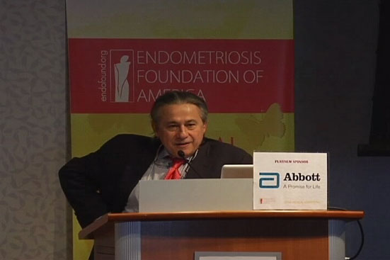
Tamer Seckin
Welcome to New York. My name is Tamer Seckin, and on behalf of the Endometriosis Foundation of America I welcome you to our first Endofound Medical Conference: Advancing the Art and Science of Endometriosis from Stem Cells to Radical Excision Surgery.
I’m so happy that I see familiar faces here. You all came from very, very far away. I know people who came from Brazil, from Puerto Rico, from Stanford and London, and since I am from Brooklyn, from the outer boroughs of Brooklyn people came here too. There are many doctors, many patients, and this shows how much you care. I congratulate you and I’m very, very happy to be here with you.
Today we are all one here, for one reason, and that is for endometriosis.
According to the latest numbers, close to 170 to 180 million people in the world, and in America alone anywhere between eight to ten million, suffer from this disease. It is the number one cause for infertility, the number one cause for pain, a major cause for hysterectomy, and a major cause for women being misdiagnosed, mistreated, and ten years of suffering and agony with late diagnosis.
We name today's session Stem Cells without discrediting the old theories. Stem cell science brings a deeper explanation and understanding to this disease. We included wide radical excision because good quality surgery happens to be the treatment that works best for these patients.
We are honored to have all of you here again for the purpose to advance the dialogue in discovery, care and treatment of women with endometriosis. I think we cannot do it alone. We need physicians, scientists, academia and advocates like you to continue to work together. With further education and awareness we can give care, relief and hope to so many millions of women in need.